Venous Ulcer of Lower Leg

Debora Sanches-Pinto, MD
, Elof Eriksson, MD

History:

66 years old woman, who has been treated for a venous ulcer in her lower leg for 4 years. She had been treated with debridement, dressing changes and compression, but the ulcer had slowly become larger. She was referred to the plastic surgery service by the vascular team. Outside of moderate hypertension, she was healthy.

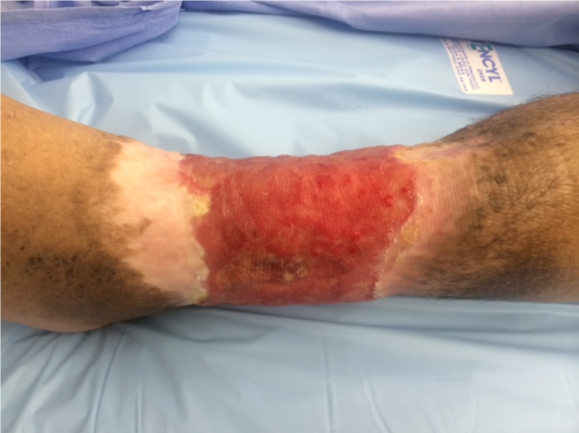
Fig.1. Ulcer at first visit.

Findings:

Ankle pulses were palpable and bounding. Ankle/Brachial Index .8. The leg had moderate to severe edema especially over the foot and a significant number of varicose veins in the leg, ankle and foot. There was a 14 X 29 cm and .4 cm deep circumferential ulcer in the lower leg. The ulcer was painful and sensitive to touch. The skin surrounding the ulcer showed dark bluish discoloration and hyperpigmentation.

Diagnosis:

Venous leg ulcer. Diagnosis was made by history and clinical examination and ulcer characteristics

Differential Diagnoses:

Mixed venous/arterial ulcer, pyoderma gangrenosum, vasculitis, cancer

Workup Required:

Gross arterial disease was ruled out. Ankle/Brachial Index as well as peripheral pulses were recorded. Color duplex ultrasound scanning with Valsalva or proximal compression for evaluation of lower leg veins was done. Deep swab culture was also done.

Plan:

Debride, reduce microbial contamination, moist dressing and compression. An ulcer of this size is very unlikely to heal with dressing changes and was scheduled for split thickness skin grafting.

Expertise Needed:

Wound Care MD, PA, NP, or RN. For Skin Grafting an MD with training in Skin Grafting is required.

Treatment:

Using local anesthesia as needed, adherent devitalized tissue was debrided using scalpel and hydro jet. The borders of ulcer with thickened, raised edges was excised. No punch biopsy to rule out malignancy was necessary. Biofilm was removed with hydro jet during debridement. An antimicrobial agent (hypochlorus acid) was then applied together with an interface dressing plus a moist foam dressing. A short stretch compression bandage was applied from the foot to the  knee. The patient was scheduled for outpatient skin grafting one week later and for another debridement in between.

One week later, the ulcer was again debrided and then micro grafted under local anesthesia as outpatient surgery. A split thickness skin graft of 25 cm square was harvested with a hand held dermatome (Xpansion, DSN, Charlestown, NH). It was then minced with a hand held mincer (Xpansion, DSN, Charlestown, NH). The minced skin particles were then spread uniformly over the wound with a spatula. The grafted area was covered with an interface dressing (Tegaderm Contact, 3M, St Paul, MN.) On top of this was placed a thin layer of hydrogel and then a “moist” foam dressing. A short stretch compression bandage was then applied. The patient was advised to keep the leg elevated.

Three, seven and ten days after surgery the patient returned for dressing changes. The hydrogel, Foam and compression bandage were replaced but the interface dressing was left in place. On postoperative day 14 and then weekly, the interface dressing was also replaced.

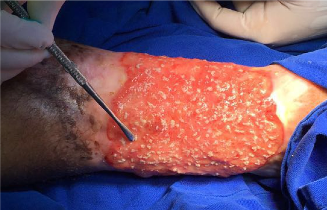
Fig.2. Ulcer with micro grafts before application of hydrogel and dressing.

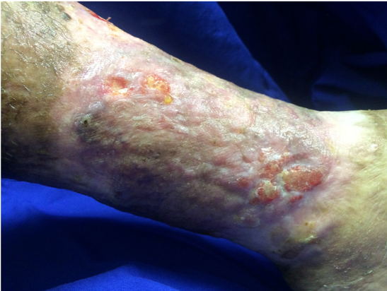
Fig.3. Grafted ulcer 3 weeks postoperatively.

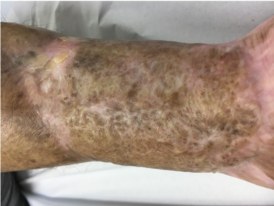
Fig.4. 8 weeks after grafting, the ulcer is almost completely healed.

Follow Up:

Three weeks after grafting, the ulcer was mainly healed. Dressing changes were then done every 2 weeks and at 8 weeks the ulcer was almost completely healed. It has remained healed for over 2 years. The patient wears a compression bandage every day.

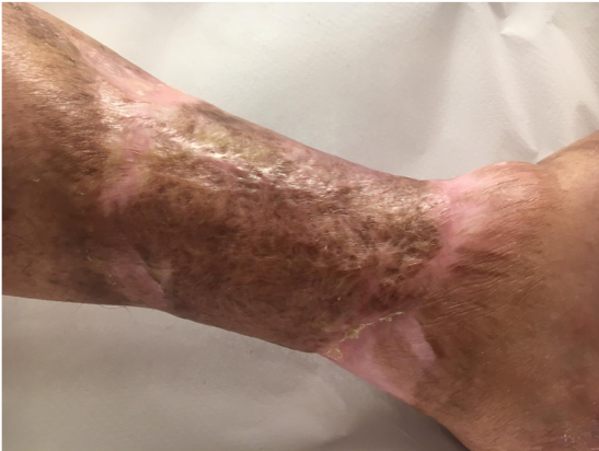
Fig.5. 2 years after grafting, the ulcer remains healed with durable skin quality.

Print Friendly, PDF & Email

References

Sieggreen,M,Y, Kline,R,A: Venous Disease, in Wound Care Essentials, edited by Baranoski,S, Ayello,E,A, 14:310, 2011.
Lisa J Gould, Garima Dosi, et al, Plast Reconstr Surg . 2016 Sep;138(3 Suppl):199S-208S. doi: 10.1097/PRS.0000000000002677.
Marston et al. Wound Healing Society 2015 update on Guidelines for Venous Ulcers. Wound Rep ,Reg, 24:136, 2016.
https://pubmed.ncbi.nlm.nih.gov/?term=Sieggreen%2CM%2CY%2C+Kline%2
https://pubmed.ncbi.nlm.nih.gov/27556762/
https://pubmed.ncbi.nlm.nih.gov/26663616/

Related Articles