Venous Ulcer Treated with Skin Graft

Rummana Aslam, MD

History:

29-year old woman presenting with a 6 X 3 cm ulcer over the left medial lower leg. It has been present for 2 years and has been treated with dressing changes but not with compression. It is painful and has slowly increased in size. She has a job requiring her to be on her feet all day. No significant medical history and no history of trauma.

Findings:

The  foot pulses are palpable and bounding. The leg has moderate to severe edema especially over the foot and a significant number of varicose veins in the leg and ankle and foot. There is a 6 X 3 cm and .4 cm deep ulcer in the medial mid lower leg. The ulcer is painful and sensitive to touch. The skin around the ulcer shows dark bluish, purple discoloration and hyperpigmentation.

Diagnosis: Venous leg ulcer. Diagnosis is made by history and clinical examination and ulcer characteristics. Differential Diagnosis:  Rule out arterial disease, mixed venous/arterial ulcer, pyoderma gangrenosum, vasculitis, cancer.

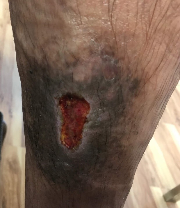
Fig. 1. 6 x 3 cm ulcer left medial lower leg

Diagnosis:

Chronic lower leg venous ulcer.

Differential Diagnoses:

Arterial ulcer.
Mixed arterial/venous ulcer
Malignant ulcer

Workup Required:

  1. Gross arterial disease was ruled out. Ankle/Brachial Index as well as peripheral pulses were recorded to be within normal limits.
  2. Color duplex ultrasound scanning with Valsalva or proximal compression showed multiple incompetent valves in both deep and superficial veins.
  3. The patient was referred  to a Vascular Surgeon for evaluation of deep and superficial veins. The incompetent deep and superficial veins were noted but no operation on the veins was recommended.
  4. Deep swab culture showing mixed skin flora and no streptococci.
  5. With no suspicion of malignancy, no biopsy for pathology was done.

Plan:

Plan: 
1. Debridement to remove indurated wound edges was done.
2. A silver/foam dressing treatment was prescribed for one week to reduce microbial contamination.
3. A moist foam dressing and a nonadherent contact layer was started after one week.
4. Compression was started with a short stretch compression bandage to reduce edema.

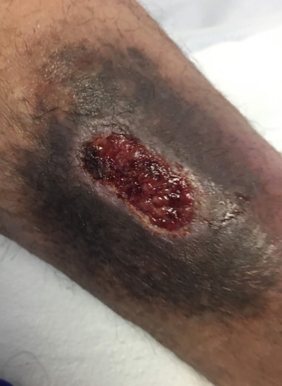
Fig.2. Ulcer after 1 week of treatment

Expertise Needed:

Wound care Practitioner – MD, PA, NP or RN

Treatment:

  1. ULCER AND SKIN CLEANSING. The ulcer was cleaned with saline to remove loose debris and slough. The skin around the ulcer should be cleaned with soap and water to reduce skin flora.
  2. DEBRIDEMENT. Under local anesthesia with .5% Lidocaine with epinephrine adherent devitalized tissue and biofilm was removed using a scalpel. The edge of ulcer which was thickened and raised was excised.
  3. INFECTION CONTROL. The wound was rinsed with hypochlorous acid and after this a silver impregnated moist foam dressing was applied. As there was no significant cellulitis and a wound culture negative for beta hemolytic streptococci; no systemic antibiotics were prescribed. .
  4. DRESSING: The contamination was greatly reduced after one week and the silver dressing was discontinued. A synthetic contact dressing plus a moist foam dressing was then applied. There was relatively little drainage so the dressing was changed every 7 days.
  5. COMPRESSION.  A short stretch compression bandage was applied to control edema and then changed with the dressing every 7 days.
  6. Split Thickness Skin Graft. After 4 weeks of treatment the ulcer was only reduced by 15% and the patient was scheduled for surgical debridement and a meshed split thickness skin graft.


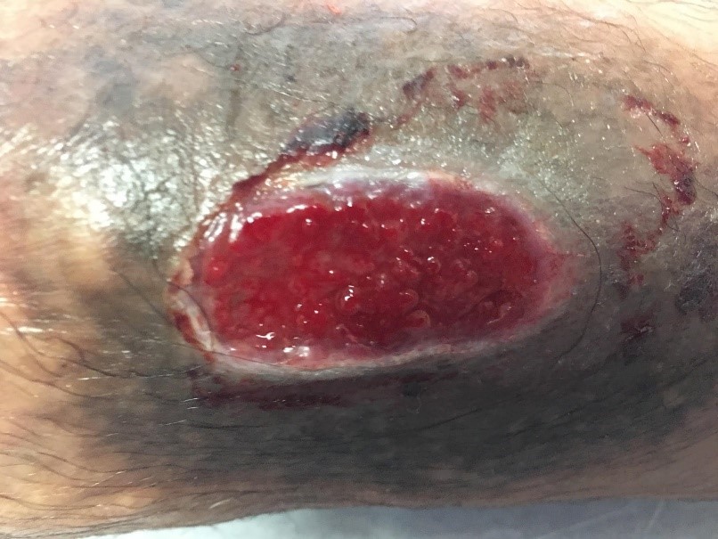
Fig.3. Ulcer after 4 weeks of treatment


Follow Up:

Two months after grafting the ulcer is completely healed.

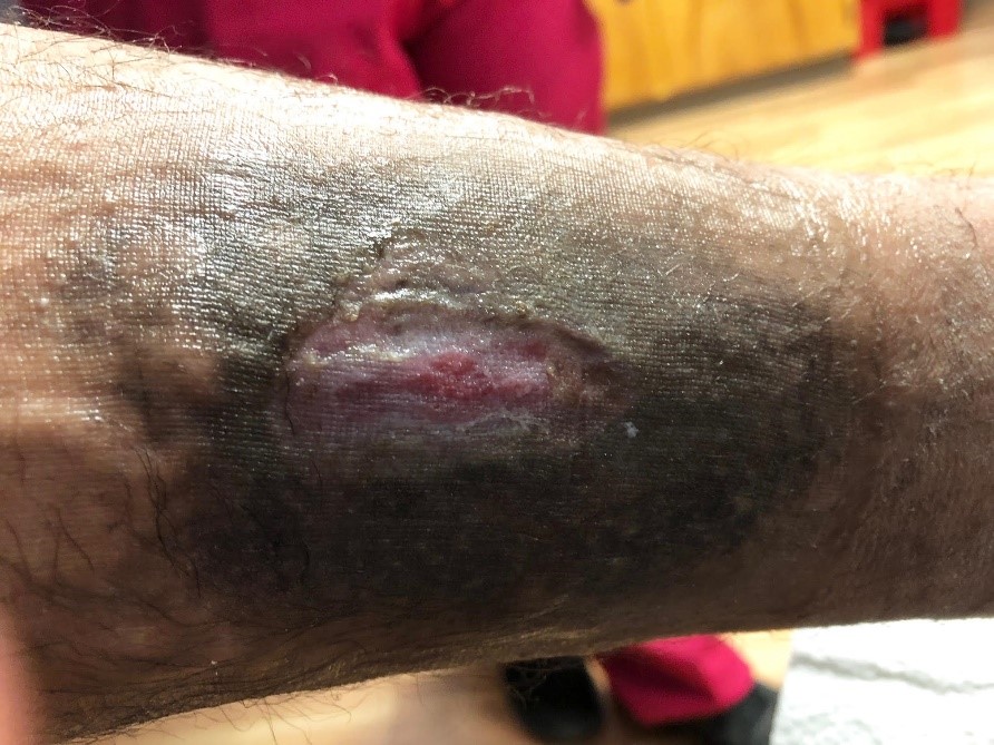
Fig.4. Healed ulcer 2 months after skin grafting


Print Friendly, PDF & Email

References

1. Harold Brem MD, Robert S Kirsner, MD, Vincent Falanga, MD. Protocol for the successful treatment of venous ulcers. The American Journal of Surgery. July 2004 Volume 188, Issue 1, Supplement 1, Pages 1–8
2. William Marston MD Jennifer Tang MD Robert S. Kirsner MD, PhD William Ennis DO, MBA. Wound healing society 2015 update on guidelines for venous ulcers. Wound Repair and Regeneration. Volume24, Issue1. January/February 2016. Pages 136-144
3. Gould, Lisa J. MD, PhD, FACS; Dosi, Garima MD, FACS; Couch, Kara MS, CRNP, CWS; Gibbons, Gary W. MD, FACS; Howell, Raelina S. MD; Brem, Harold MD, FACS; Tomic-Canic, Marjana PhD Modalities to Treat Venous Ulcers: Compression, Surgery, and Bioengineered Tissue. Plastic and Reconstructive Surgery: September 2016 - Volume 138 - Issue 3S - p 199S–208S
https://www.americanjournalofsurgery.com/article/S0002-9610(03)00284-8/fulltext
https://pubmed.ncbi.nlm.nih.gov/26663616/
https://pubmed.ncbi.nlm.nih.gov/27556762/

Related Articles