Upper eyelid laceration in multi-trauma patient

Olivier F. Noel, M.D., Ph.D.
, Jackie Oh, M.D.
, Omar Allam, M.D.

History:

A 22 year old who presented after a motorcycle accident. He had not been wearing a helmet and had loss consciousness at the scene where he was intubated by EMS. He suffered several other injuries including splenic laceration which necessitated an exploratory laparotomy and a left upper eyelid deep laceration. CT scans of head and abdomen were obtained upon arrival to the ED. Monitoring of respiratory and hemodynamic parameters was started upon arrival to the ED. Plastic Surgery was consulted to repair his eyelid laceration.

Findings:

On exam, 2.5 cm deep laceration extending into the orbicularis muscle and down to the lateral orbital rim. The eyelid has some erythema and edema. We were unable to assess the patient’s visual acuity due to his condition. There was no loss of teeth or evidence of facial fractures.

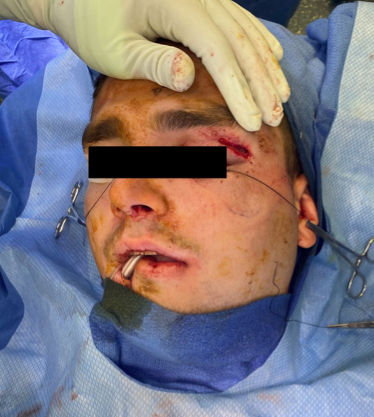
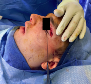
Figure 1-2. Show left eyelid laceration.

Diagnosis:

Complex laceration of left upper eyelid. No orbital or other facial fractures.

Differential Diagnoses:

R/O ocular injury.

Workup Required:

ACLS trauma work up performed in the ED. No further workup required from the laceration standpoint. Ophthalmology consultation was requested.

Plan:

Repair of the laceration while the patient was intubated under anesthesia in the Operating Room.

Expertise Needed:

Plastic surgeon.

Treatment:

Wound gently cleaned with saline and betadine by gentle dabbing with dry sterile gauze. In the muscle, 5-0 Monocryl sutures were used for orbicularis muscle approximation. 6-0 Monocryl dermal sutures were used to approximate the skin edges. 6-0 Prolene interrupted sutures were used for the best skin approximation. Bacitracin was applied to the wound and the patient’s caregivers were instructed to apply Bacitracin ointment twice daily until follow up. He was also given written instructions to avoid sun exposure and use SPF-30 or higher lotion if exposed to sun for the next year. This was done in order to decrease the risk of hyper pigmentation of her scar. He was scheduled for follow up in 1 week pending discharge for suture removal. He received 3 days of prophylactic antibiotics.

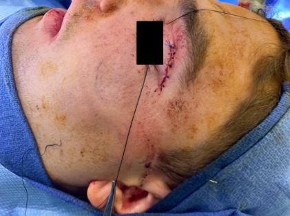
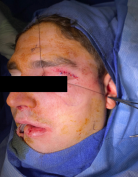
Figure 3-4. Show repaired eyelid laceration.

Follow Up:

The patient was hospitalized and the wound dressing was changed daily for 4 days. The patient was discharged on day 13 after the operation without any wound complications. At the follow up after 21 days the wound showed good healing without complications.

Print Friendly, PDF & Email

References

Franz MG, Robson MC, Steed DL, Barbul A, Brem H, Cooper DM, Leaper D, Milner SM, Payne WG, Wachtel TL, Wiersema-Bryant L; Wound Healing Society. Guidelines to aid healing of acute wounds by decreasing impediments of healing. Wound Repair Regen. 2008 Nov-Dec;16(6):723-48. doi: 10.1111/j.1524-475X.2008.00427.x. PMID: 19128244.
Bhattacharya V. Management of soft tissue wounds of the face. Indian J Plast Surg. 2012 Sep;45(3):436-43. doi: 10.4103/0970-0358.105936. PMID: 23450264; PMCID: PMC3580340.
Cochran ML, Czyz CN. Eyelid Laceration. 2022 Oct 7. In: StatPearls [Internet]. Treasure Island (FL): StatPearls Publishing; 2022 Jan–. PMID: 29261910.
https://pubmed.ncbi.nlm.nih.gov/19128244/
https://www.ncbi.nlm.nih.gov/pmc/articles/PMC3580340/
https://pubmed.ncbi.nlm.nih.gov/29261910/

Related Articles