Trochanteric Pressure Sore in Patient with advanced MS closed with Tensor Fascia Lata FlapS

Elof Eriksson, MD

History:

52 year old patient with a 5 year history of MS that has progressed in spite of active treatment. He is bedridden since 2 years and has developed hip and knee flexion contractures preventing him from lying on his back or abdomen. As a result he has developed bilateral trochanteric pressure sores. Offloading, soft mattresses and pads have had no effect. The larger right pressure sore has been present for over a year. It has been treated by his primary care physician and in spite of optimal treatment slowly become larger. He is referred to plastic surgery for surgical treatment of his pressure sore.

Findings:

52 year old man who is alert and oriented. He is bedridden. Over the right trochanter, he has an 8 X 8 cm deep ulcer. It extends down to and around the trochanter and is covered by healthy granulation tissue. There is a small amount of skin necrosis in the periphery of the ulcer. There is no evidence of active infection in the ulcer. The patient is taking a number df medications for his MS as well as for muscle spasms.
The patient also has a left trochanteric pressure ulcer which will be addressed later.

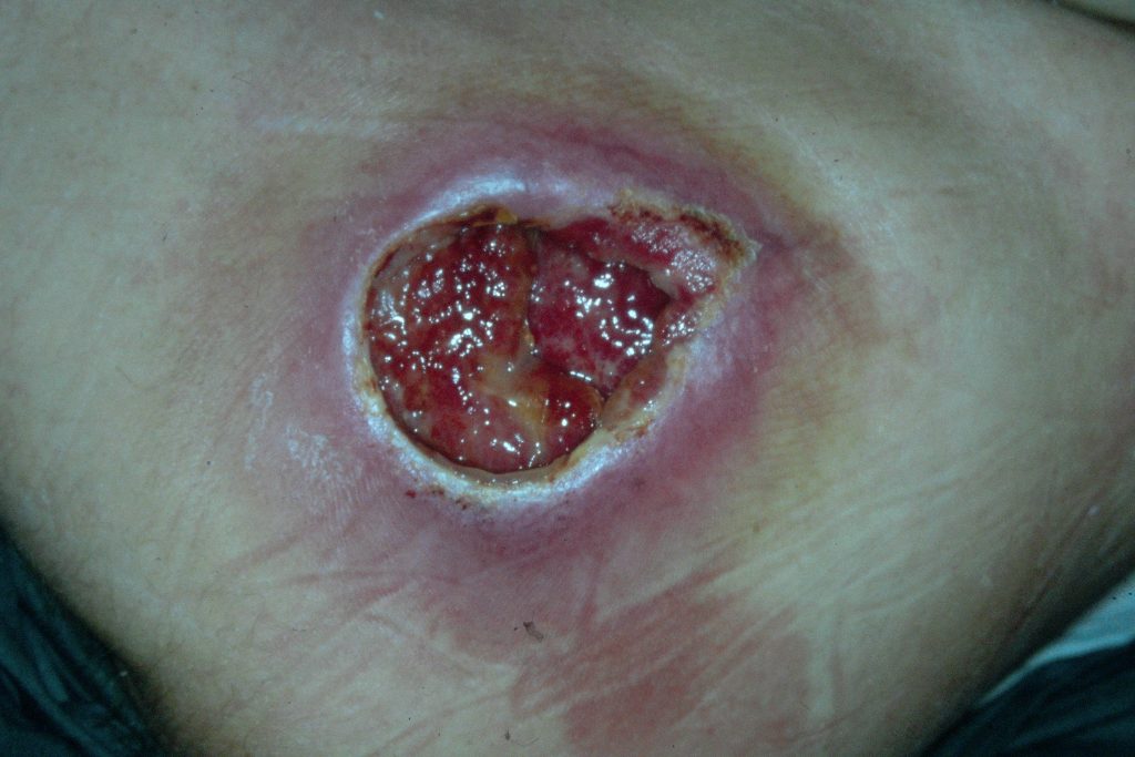
Fig. 1. Right trochanteric pressure sore.

Diagnosis:

Right trochanteric pressure ulcer extending to and around most of the trochanter. The bone of the greater trochanter is exposed and devoid of periosteum in the most prominent portion. No evidence of invasive. infection.
Left trochanteric pressure ulcer which will not be addressed at this admission.

Differential Diagnoses:

None

Workup Required:

Wound culture, x-ray of hip to assess bone destruction, it was decided not to do CT-scan, MRI or bone biopsy because of likelihood of low additional yield,, laboratory studies including full protein profile and hemoglobin A1C. Full physical examination. Consult with MS specialist.

Plan:

Hospital admission with debridement of ulcer with flap closure.

Expertise Needed:

Plastic Surgeon with experience in Pressure Sore treatment.

Treatment:

The workup was done as an outpatient. The patient was admitted on the day of surgery. Under general anesthesia the whole ulcer cavity was stained with Methylene Blue and then excised in a ‘pseudotumor’ fashion. The prominent part of the exposed greater trochanter (2 cm thick in its thickest portion) was removed with a chisel. After careful hemostasis, a Tensor Fascia Lata Flap was dissected and transposed to cover the defect. The flap donor site was covered with a split thickness skin graft from the same anterior thigh. The patient was transferred to a Rehab Hospital 7 days later. At that time, the flap and the skin graft were healing without any problems.

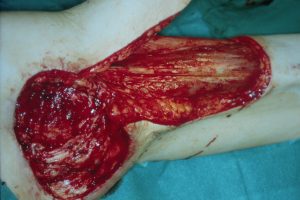
Fig. 2. Trochanteric pressure sore resected and tensor fascia lata flap elevated.

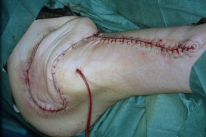
Fig. 3. Flap sutured in place.

Follow Up:

One month after his operation, the patient was completely healed and was scheduled for surgery on the trochanteric pressure sore on the opposite hip.

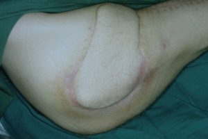
Fig.4. The healed wound 2 months later.

Print Friendly, PDF & Email

References

Multiple Flaps for Trochanteric Pressure Sore Reconstruction: A Case Series
Free flaps for pressure sore coverage
Multi-disciplinary management of complex pressure sore reconstruction: 5-year review of experience in a spinal injuries centre
https://pubmed.ncbi.nlm.nih/32850237
https://pubmed.ncbi.nlm.nih/18520197
https://pubmed.ncbi.nlm.nih/27490980

Related Articles