
Traumatic Ear Laceration

Zohra Aslami, MD
, Indranil Sinha, MD

History:

We present the case of a 43-year-old-male, with a past medical history of left leg deep vein thrombosis and Factor V Leiden on Eliquis, who presented to the emergency department after a vasovagal episode in his home resulting in an unwitnessed fall in his kitchen.

His ear struck the kitchen counter as he was falling, resulting in a laceration. In the emergency room he was hemodynamically normal, laboratory findings were reassuring, and a CT Head did not demonstrate any acute findings.

Findings:

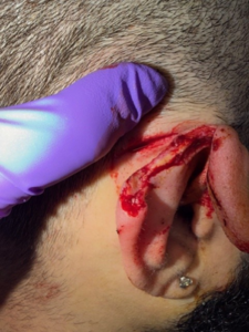
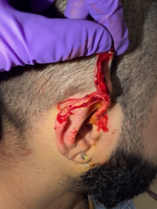
During physical examination, the patient was found to have a full thickness ear laceration through the superior helix and scapha. Cartilage was exposed and there was a sub-centimeter section of soft tissue attaching the lacerated portion to the helical root. There was an associated postauricular linear laceration, with depth to subcutaneous tissue. The initial appearance was photographed as pictured
Diagnosis:

Full thickness linear ear laceration through superior helix and scapha

Differential Diagnoses:

None

Workup Required:

Appropriate TDAP and ancef prophylaxis should be provided if indicated. Any indicated trauma or syncope workup e.g. CT Head should be performed if appropriate given the mechanism of injury. A thorough history including allergies, bleeding or clotting disorders, and wound healing difficulties should be assessed, as well as a full craniofacial examination to rule out other injuries.

Plan:

Bedside laceration repair under local anesthesia

Expertise Needed:

Plastic Surgeon, Craniofacial Surgeon, Ear Nose Throat Surgeon, or Oral Maxillofacial Surgeon

Treatment:

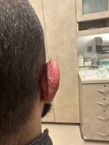
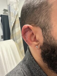
In this case, local anesthesia was obtained via local infiltration of 1% lidocaine with epinephrine via auricular ring block. The laceration was thoroughly irrigated with dilute betadine and saline. The fractured cartilage was re- approximated with 5-0 monocryl suture, while the skin was approximated with 5-0 plain gut suture. The associated postauricular laceration was repaired in layers with 5-0 monocryl deep dermal interrupted sutures and interrupted 5-0 plain gut sutures for the skin. A xeroform bolster was fashioned and sutured in with a 3-0 prolene mattress suture. A 5-day prophylactic ciprofloxacin course was prescribed given exposure of cartilage at time of injury.

Follow Up:

The patient was seen in follow up 5 days after the repair for bolster removal and the incision was healing well at the time with no associated hematoma or chondritis.
Print Friendly, PDF & Email

References

Williams CH, Sternard BT. Complex ear lacerations. In: StatPearls. Treasure Island, FL: StatPearls Publishing; 2023. Accessed July 31, 2023. https://pubmed.ncbi.nlm.nih.gov/30247848/
Kaplan AL, Cook JL. The incidences of chondritis and perichondritis associated with the surgical manipulation of auricular cartilage. Dermatol Surg. 2004;30(1):58-62.
Dalal PJ, Purkey MR, Price CPE, Sidle DM. Risk factors for auricular hematoma and recurrence after drainage. Laryngoscope. 2020;130(3):628-631. doi:10.1002/lary.28310
https://pubmed.ncbi.nlm.nih.gov/30247848/
https://pubmed.ncbi.nlm.nih.gov/14692929/
https://pubmed.ncbi.nlm.nih.gov/31621925/

Related Articles
