Temporal Fossa Defect Repair After Craniectomy

Elise Hogan, BS
, Ryan Gobble, MD

History:

A 51-year-old Caucasian female with a history of a left sphenoid wing meningioma s/p craniectomy and resection presented to plastic surgery clinic four years later with desire for repair of temporal defect. The patient also experienced dehiscence of the left levator palpebrae muscle, leading to ptosis and visual impairment. She was additionally bothered by excess skin of her upper eyelids.

Findings:

On exam, patient was noted to have a left temporal fossa contour abnormality measuring 4x4cm with depression as seen in Figure 1, as well as unilateral ptosis and vertical dystopia. She had excess skin to the bilateral upper eyelids with drooping.

Figure 1: Frontal, anterolateral, and lateral profiles showing left temporal defect, left ptosis, and excess eyelid skin bilaterally.

Diagnosis:

    1. Left temporal hollowing
    2. Left levator palpebrae muscle dehiscence
    3. Excessive upper eyelid skin bilaterally

Differential Diagnoses:

None

Workup Required:

No additional workup was required.

Plan:

To address the temporal hollowing, with structural fat grafting from the abdomen was recommended to restore normal temporal contour. Risks such as intra-abdominal injury from liposuction cannula, contour irregularity or grooving from liposuction, and fat necrosis as well as the need for future procedures were discussed and the patient elected to move forward with treatment.

To address the patient’s ocular complaints, she would first undergo visual field testing in order to submit for insurance coverage. For levator palpebrae dehiscence, muscular re-suspension was planned.  In order to address the patients concerns for excess skin of the upper eyelids, a bilateral blepharoplasty was planned.

Expertise Needed:

A plastic surgeon with expertise in soft tissue injury was required.

Treatment:

To address the temporal fossa defect, the abdomen was prepped and fat was collected via 4mm Mercedes tip liposuction cannula, and the incisions closed with 5-0 fast gut.

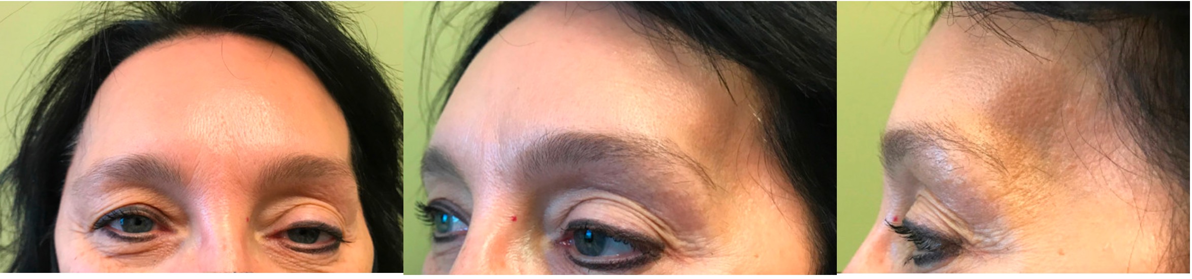
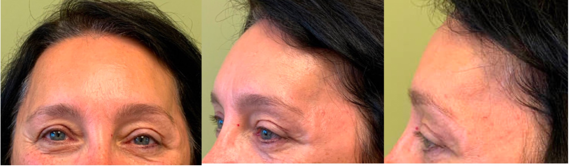
The upper blepharoplasty was performed next. A caliper was used to mark the lower border of the incision and approximately 8-10 mm throughout the length of the lower incision above the eyelid margin. A pinch test was then performed to determine the superior extent of the skin excision, and the skin was then injected with local anesthetic (lidocaine 1% with 1:100,000 epi). The skin was then excised using a beaver blade bilaterally. A thin strip of orbicularis oculi muscle was then excised using black handle scissors, exposing the the orbital septum bilaterally. On the left side, the obicularis muscle was dissected from the anterior aspect of the tarsus and levator aponeurosis. The levator palpebrae was predominantly still attached; however, was quite stretched and attenuated along with an element of dehiscence and at this point, the levator muscle was resuspended. This was done carefully and by observing the other eye, to maintain symmetry. Once satisfied, the levator aponeurosis-Muller’s muscle complex was reinserted into the anterior aspect of the tarsal plate using a 4-0 Monocryl. Next both the right and left orbicularis oculi muscles were reapproximated using 4-0 Monocryl ,and the incisions were closed with a running 4-0 prolene. Laterally, 3 inturrupted sutures were placed on each side.

Finally, attention was turned to the left temporal area. 3 small stab incisions were made through which the previously harvested fat was injected using a micro aliquot technique and only while withdrawing the Coleman injection canula. A total of 9mL was placed, which resulted in a bilaterally symmetric appearance. Once this was completed, the small stab incisions were closed with a 5-0 fast gut and covered with dermabond. The eyelid incisions were treated with ophthalmic bacitracin. The eyelids were then covered with ice water soaked 4×4 gauze, making sure not to allow this to sit over the temporal area where the fat was grafted.

Follow Up:

The patient was seen two months later with no complications. She has well healed blepharoplasty scars with correction of left upper eyelid ptosis. There was improvement in contour of the left temporal region; however, there was still a slight depression relative to the right side which could be improved with another round of fat grafting. The patient expresses interest in this and will follow up when she decides to proceed. The vertical dystopia remained post-operatively.

Figure 2: Frontal, anterolateral, and lateral profiles post operatively showing improvement in symmetry of the temporal fossa, ptosis, and decreased upper eyelid skin.

Print Friendly, PDF & Email

References

1. Finsterer J. (2003). Ptosis: causes, presentation, and management. Aesthetic plastic surgery, 27(3), 193–204.
2. Franz, M. G., Robson, M. C., Steed, D. L., Barbul, A., Brem, H., Cooper, D. M., Leaper, D., Milner, S. M., Payne, W. G., Wachtel, T. L., Wiersema-Bryant, L., & Wound Healing Society (2008). Guidelines to aid healing of acute wounds by decreasing impediments of healing. Wound repair and regeneration : official publication of the Wound Healing Society [and] the European Tissue Repair Society, 16(6), 723–748.
3. Kaufman, M. R., Miller, T. A., Huang, C., Roostaien, J., Wasson, K. L., Ashley, R. K., & Bradley, J. P. (2007). Autologous fat transfer for facial recontouring: is there science behind the art?. Plastic and reconstructive surgery, 119(7), 2287–2296.
https://doi.org/10.1007/s00266-003-0127-5
https://doi.org/10.1111/j.1524-475X.2008.00427.x
https://doi.org/10.1097/01.prs.0000260712.44089.e7

Related Articles