Stellate Lip and Cheek Laceration

Mariana Trevino, MD
, Deepak Bharadia, MD
, Indranil Sinha, MD

History:

A healthy 3-year-old boy presents to the outside hospital emergency room 5 hours after a dog bite to the face. Patient did not lose consciousness and there were no other injuries noted. He presented with pain at the site of the laceration and minimal bleeding. Physical examination revealed a complex, stellate laceration on the left side of the face. Patient received intravenous Augmentin prior to transfer and immunizations were up to date. There were no abuse/neglect concerns.

Findings:

5×4 cm sized full-thickness complex laceration of skin with avulsed tissue involving vermillion border of both upper and lower lip and commissure on left side.  The laceration extended to adjacent cheek with exposed subcutaneous tissue.  There is no intra-oral mucosal injury present.The patient appeared to have some motion in both lateral upper and lower lip.

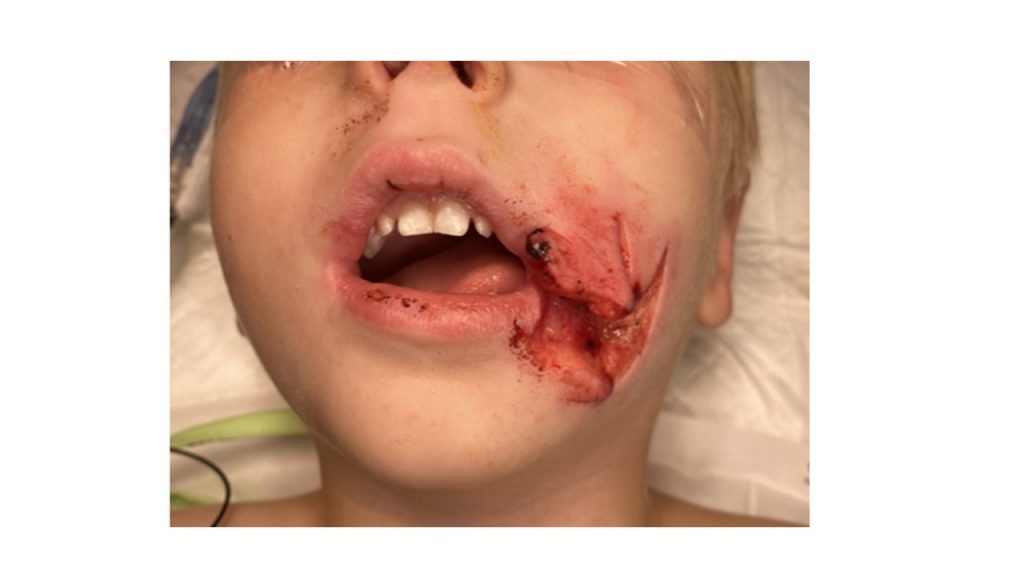
Figure 1. Patient presents with stellate cheek laceration involving his left upper lip.

Diagnosis: 5 cm x 4 cm complex laceration involving vermillion border and adjacent cheek and with tissue loss in the lateral lower lip. No involvement of facial nerve branches or Parotid duct.

Differential Diagnosis: None

Work-Up Required: A complete physical exam was performed, including testing of mouth opening and any bony step-offs (1).  As part of a physical exam, facial nerve function was systematically tested by evaluating motion at site of laceration (2).  Motion was limited by pain.  Evaluation of parotid (Stensen’s) duct under general anesthesia was done  by insertion of an angiocath canula into the intra oral opening of the parotid duct and injection of saline, to detect fluid extravasation into the cheek.

Plan: Washout and complex laceration repair under local anesthesia with sedation in Emergency Room or Operating Room.

Expertise Needed: Plastic Surgery consultation. Sedation could not be performed reliably in the Emergency Room,  and Plastic Surgery  repair was performed in the Operating Room, under general anesthesia.

Treatment: The patient was given Ketamine for anesthesia. His face was then prepped and draped in usual sterile fashion using betadine. Methylene blue was used to outline the vermillion border. Small areas of denuded tissue were then debrided prior to approximating the skin. No muscles had been severed. The deep dermal layer was approximated using 4-0 and 5-0 Monocryl sutures and the skin was closed using 5-0 fast absorbable gut in an interrupted fashion. Bacitracin and Telfa and Tegaderm were applied over the repaired lacerations.

Figure 2. After irrigation and cautious debridement, the tissues were reapproximated and secured with sutures in the operating room.

Follow up:  The patient was prescribed Augmentin for one week.  The parents were asked to cover the laceration with bacitracin ointment for the first week and the patient could resume facial washing 48 hours after repair.  Tylenol and Motrin was recommended for pain control.  Follow up in 1 week for evaluation and removal of any sutures which had not dissolved. Recommended application of sunscreen to laceration site, starting at 2 weeks following repair, and every 2 hours when in sunlight for at least 1 year to minimize scar conspicuousness (3).

Figure 3A. Patient demonstrates a well healing wound without functional impairment.

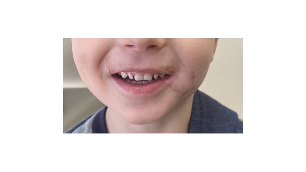
Figure 3B. At the last follow up the patient demonstrated a well healing wound without functional impairment.


Print Friendly, PDF & Email

References

Hwang K. Field Management of Facial Injuries in Sports. J Craniofac Surg. 2020 Mar/Apr;31(2):e179-e182.
Rogan DT, Fang A. Pediatric Facial Trauma. 2020 Jul 6. In: StatPearls [Internet]. Treasure Island (FL): StatPearls Publishing; 2021 Jan–. PMID: 32644358.
Shimizu I, MacFarlane D. Zinc oxide paste as sunscreen in the postoperative period. Dermatol Surg. 2012 Jun;38(6):965-6
https://pubmed.ncbi.nlm.nih.gov/31895853/
https://pubmed.ncbi.nlm.nih.gov/32644358/
https://pubmed.ncbi.nlm.nih.gov/22642456/

Related Articles