Stab Wound to Forearm

SeungJu Jackie Oh MD
, Olivier Noel MD
, Omar Allam MD
, Adnan Prsic MD

History:

40-year-old right-handed, otherwise healthy male patient who presented as a full trauma activation patient after sustaining multiple stab wounds to left chest and upper extremity during a robbery.

Findings:

8-cm laceration to left volar forearm. Partial transection of FCR (Flexor Carpi Radialis) muscle belly. Motor and sensation fully intact. Warm and well-perfused, vascularly intact with capillary refill <3 seconds in all finger tips. Palpable radial and ulnar pulse. Flexion/extension intact in wrist. Flexion of FDP/FDS and extension intact in all fingers. Median/radial and ulnar motor function grossly intact with “OK” sign, thumbs up and finger adduction/abduction. Sensation intact in radial, median and ulnar distribution.

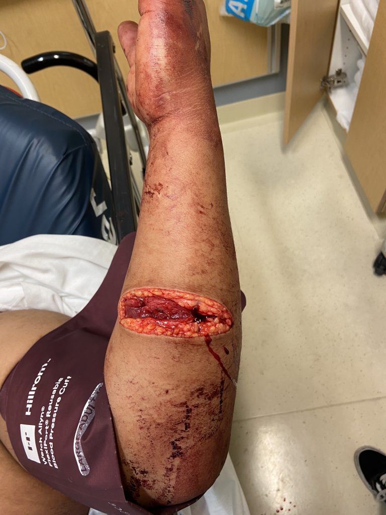
Figure 1: Lateral view- 8cm laceration to left volar forearm. Partial transection of FCR muscle belly.

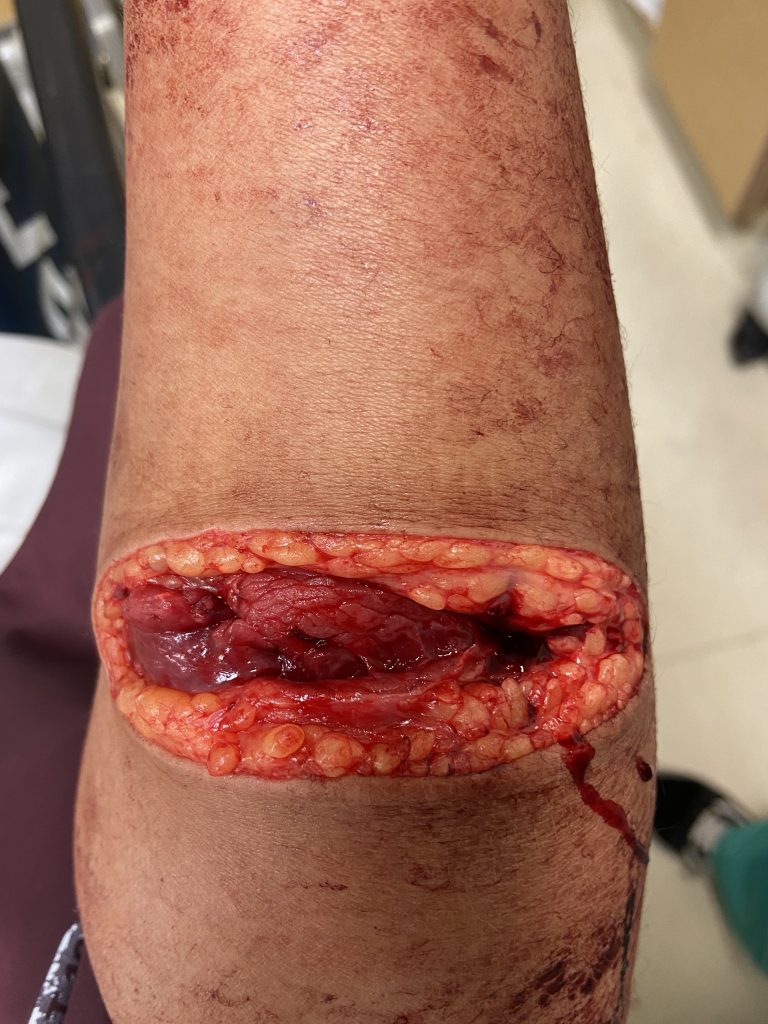
Figure 2: Anterior view of left volar forearm laceration.

Diagnosis:

Deep laceration to left volar forearm involving flexor carpi radialis muscle belly

Differential Diagnoses:

R/O neuro-vascular injuries.

Workup Required:

Full ATLS work-up. In the trauma bay, primary survey intact and hemodynamically stable. GCS 15 (eye, verbal, and motor response. 15 equals normal). Secondary survey notable for 10-cm laceration over left lateral chest and deep 8-cm laceration to left forearm. Initially hypotensive, so received aggressive fluid resuscitation with appropriate response. CT head, c-spine, chest, abdomen and pelvis negative for other acute traumatic injuries. Left upper extremity XR (humerus, elbow, forearm and wrist) also negative for bony injuries. Palpable radial and ulnar pulses, low clinical concern for vascular injury. After initial management, was admitted to trauma surgery service.

Plan:

Washout and laceration repair in ED.

Expertise Needed:

Plastic or Hand Surgeon.

Treatment:

Local field block was achieved with 20cc of 1% lidocaine with epinephrine. Wound was thoroughly irrigated with 1L NS mixed with Betadine. FCR muscle belly and muscle fascia approximated with 3-0 Vicryl sutures. Dermis was loosely approximated with 3-0 Monocryl (buried dermal sutures) and skin with 4-0 Chromic Gut (simple interrupted). Wound then dressed with bacitracin, xeroform, kerlix and ACE wrap. Patient tolerated the procedure well.

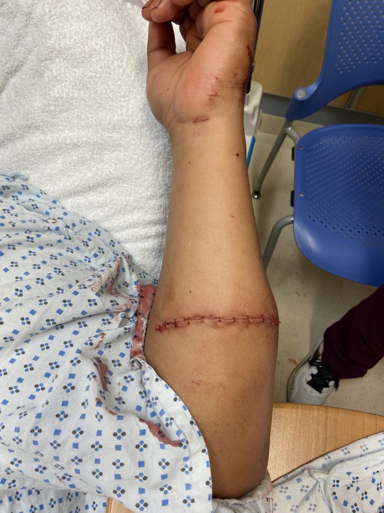
Figure 3: S/p layered closure of left volar forearm laceration.

-Discharge Instructions:

  • Keflex for 1 week
  • Keep dressing on for 2 days, followed by daily dressing changes with bacitracin/ xeroform/ kerlix and ACE wrap
  • LUE elevation as much as possible
  • Follow-up PRS clinic 1 week for wound check

Follow Up:

Well-healing wound at 2-week clinic visit (image below). Dressing changed to daily bacitracin and left open to air. Patient seen once more at 1-month post-injury, and was cleared to follow-up on prn basis.

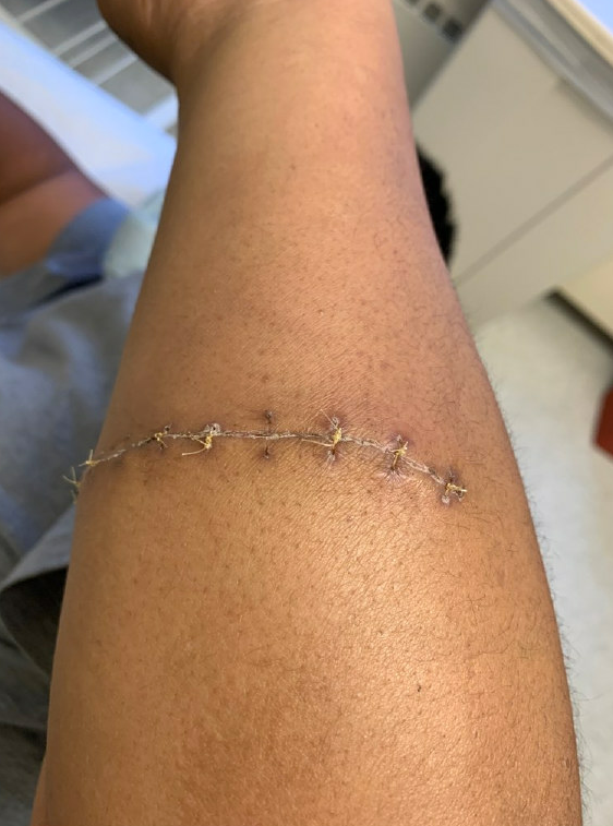
Figure 4: Well-healing wound 2 weeks s/p repair.

Print Friendly, PDF & Email

References

Bashir, Muhammad Mustehsan, Muhammad Sohail, and Hussan Birkhez Shami. "Traumatic wounds of the upper extremity: coverage strategies." Hand clinics 34.1 (2018): 61-74.
Wolfe, Scott W., et al. Green's operative hand surgery. Elsevier Health Sciences, 2021.
Daniels, James M., Elvin G. Zook, and James M. Lynch. "Hand and wrist injuries: Part II. Emergent evaluation." American Family Physician 69.8 (2004): 1949-1956.
https://pubmed.ncbi.nlm.nih.gov/29169598/
https://www.elsevier.com/books/greens-operative-hand-surgery/bresnahan/978-0-323-69793-4
https://www.aafp.org/pubs/afp/issues/2004/0415/p1949.html

Related Articles