Pyoderma Gangrenosum of Finger

Samantha Lund, MD
, Nada Berry, MD
, Nicole Sommer, MD

History:

79-year-old woman with past medical history of diabetes, hypertension, atrial fibrillation, and transient ischemic attack first presented to an outside hospital emergency room with a spontaneous blister over her left index finger dorsal-radial proximal interphalangeal joint. She denied trauma to the area. This was aspirated and she was prescribed oral antibiotics. She followed up with her primary care physician and reported further progression of the wound to ulceration. Hematoma with purulence was aspirated; the fluid resulted in a negative culture. She was then referred to a hand surgeon who excised a 2x1cm caseating, friable lesion that was sent to pathology to ultimately show nonspecific inflammation. At the recommendation of the hand surgeon, she presented to the emergency room for admission and workup for pyoderma gangrenosum.

Findings:

At the time of presentation to the emergency room, the wound had been present for approximately two weeks with continued progression despite antibiotics, aspiration, and excision debridement. The wound had expanded to encompass the radial side of the digit with violaceous bullae with central ulceration. It was very painful for the patient. With the history and exam, there was concern for pyoderma gangrenosum.

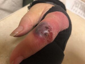
Initial presentation to PCP
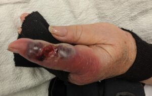
Presentation to ED and evaluation by hand surgery

Diagnosis:

The primary care physician and plastic surgeon workup included a negative wound culture with nonspecific inflammation on pathology. Dermatology conducted a series of lab tests that revealed mild leukocytosis with mild elevation of CRP. ANA was positive with a titer of 1:80 and a homogenous pattern. RF was negative, SPEP/UPEP normal, ANCA negative, and a negative fecal occult blood test. The patient denied exposure to gardening, fish, or other animals/insects. She denied gastrointestinal symptoms. She had no history of skin cancer or other dermatologic or rheumatologic diseases.

Pyoderma gangrenosum can be associated with underlying autoimmune conditions, such as irritable bowel disease and rheumatoid arthritis. However, the patient’s history was negative for other conditions and the only positive lab result was the low titer and homogenous pattern of the ANA test. Thus, the diagnosis of idiopathic bullous pyoderma gangrenosum was made.

Differential Diagnoses:

  • Pyoderma gangrenosum: diagnosis of exclusion, positive ANA, improved with steroid treatment
  • Soft tissue infection: negative culture and pathology, mild leukocytosis, no inciting injury
  • Vasculitis: negative pathology, no new medication, no prior illness, negative ANCA
  • Skin cancer: negative pathology
  • Sporotrichosis: no exposure to gardening
  • Vibrio vulnificus: no exposure to fresh water or fish

Workup Required:

Thorough history, wound culture, tissue pathology, autoimmune laboratory tests

Plan:

Dermatologic assessment 

Expertise Needed:

Dermatology, plastic surgery, hand surgery

Treatment:

The patient was started on oral prednisone with topical clobetasol daily dressings. The initial dose was 60mg which had an immediate effect on alleviating the patient’s pain. The dose was decreased to 40mg and subsequently tapered over two months.

Follow Up:

The patient followed up with dermatology and with plastic surgery. One week after prednisone treatment was initiated, the wound was healing with resolution of pain. Three weeks later the wound had completely resolved. The wound has not recurred in six months of follow up.

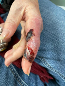
First follow up visit at one week after hospitalization
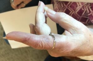
Six month follow up
Print Friendly, PDF & Email

References

Untreatable surgical site infection: consider pyoderma gangrenosum
Pyoderma gangrenosum. A comparison of typical and atypical forms with an emphasis on time to remission. Case review of 86 patients from 2 institutions
The histological assessment of cutaneous vasculitis
https://www.ncbi.nlm.nih.gov/pmc/articles/PMC4174238/
https://pubmed.ncbi.nlm.nih.gov/10670408/
https://pubmed.ncbi.nlm.nih.gov/20055902/

Related Articles