Pediatric Subungual Hematoma and Nail Bed Laceration

SeungJu Jackie Oh MD
, David Colen MD

History:

8-year-old right-handed female patient presenting with right ring finger injury after door slammed shut on finger two days ago. Given worsening swelling and pain, mother decided to present to ED for further evaluation.

Findings:

Significant subungual hematoma limited to dorsal aspect of right RF distal phalanx. Digit otherwise warm and well-perfused, vascularly intact with capillary refill <3 seconds in all finger tips. Palpable radial and ulnar pulse. Flexion/extension intact in wrist. Flexion of FDP/FDS and extension intact in all fingers. Median/radial and ulnar motor function grossly intact with “OK” sign, thumbs up and finger adduction/abduction. Sensation intact in radial, median and ulnar distribution.

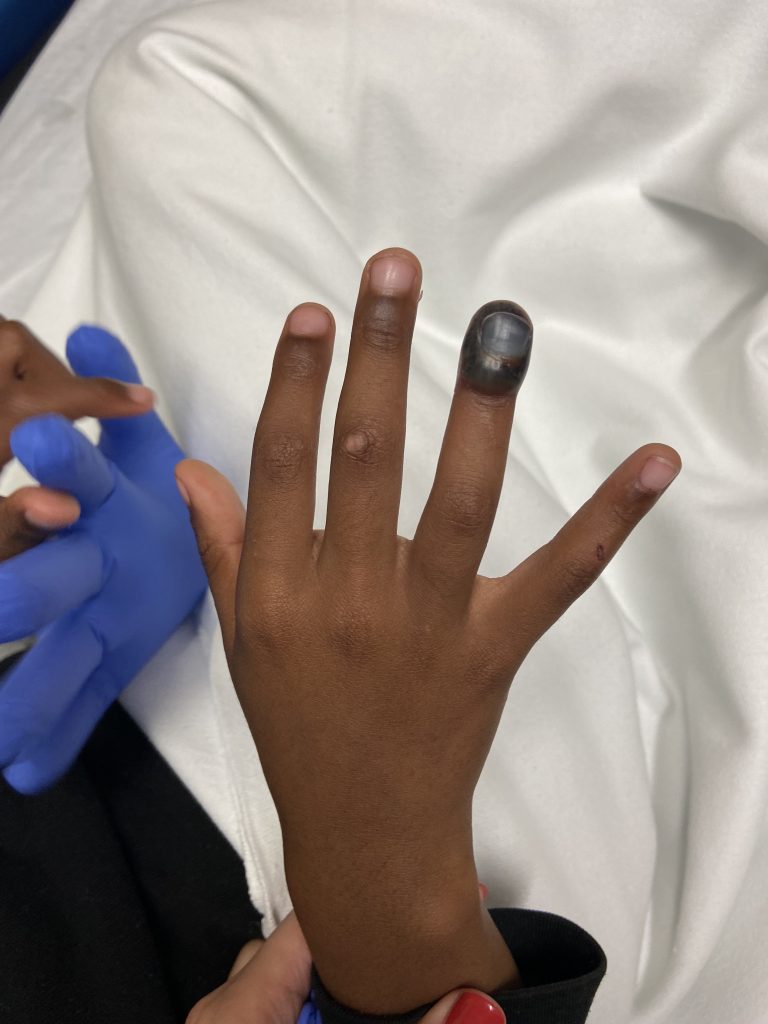
Figure 1: Anterior view of the subungual hematoma of right RF.

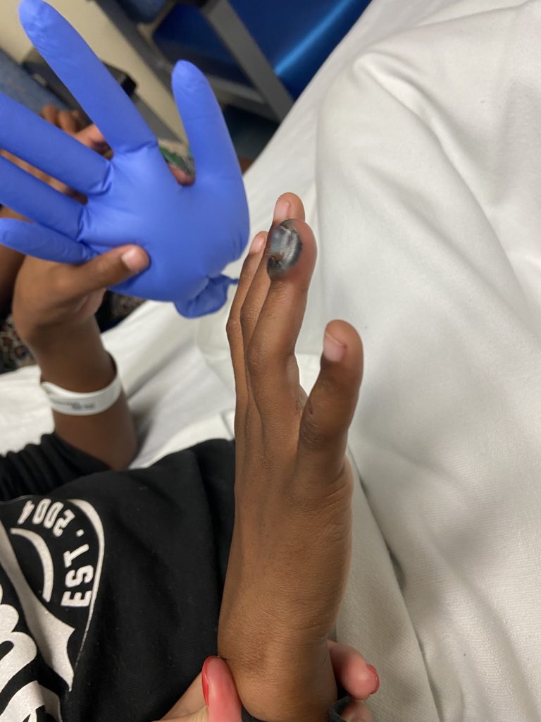
Figure 2: Lateral view of subungual hematoma limited to dorsal aspect of right RF distal phalanx.

Diagnosis:

Right ring finger subungual hematoma with likely nail bed laceration (especially given hematoma >50% of nail).

Differential Diagnoses:

R/O Seymour fracture (displaced distal phalanx physeal fracture with associated nail bed laceration)

Workup Required:

Complete hand exam (as detailed above). Hand and isolated finger XR to rule out Seymour fracture. Xray ruled out Seymour fracture.

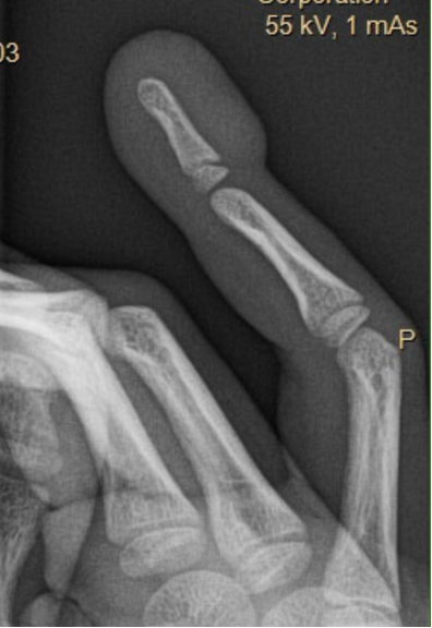
Figure 3: Dedicated right RF finger X-ray (lateral view) without Seymour fracture.

Plan:

Wash out, nail removal, nail bed laceration repair in ED.

Expertise Needed:

Plastic or Hand surgeon.

Treatment:

Intra-nasal versed and digital block using 6cc of 1% lidocaine without epinephrine. Nail was removed using a periosteal elevator with immediate drainage of 10cc of blood. Visible nail bed laceration distal to the nail fold. No instability noted of the distal phalanx. Wound thoroughly irrigated with 250cc NS mixed with betadine. Nail bed lac repaired with 5-0 fast gut. Stented nail with original nail which was secured with dermabond. Dressed with bacitracin, xeroform gauze, and kling wrap. Alumafoam splint was applied to prevent accidental trauma to the fingertip and for patient comfort. Patient tolerated the procedure well.

Visible nail bed laceration distal to nail fold

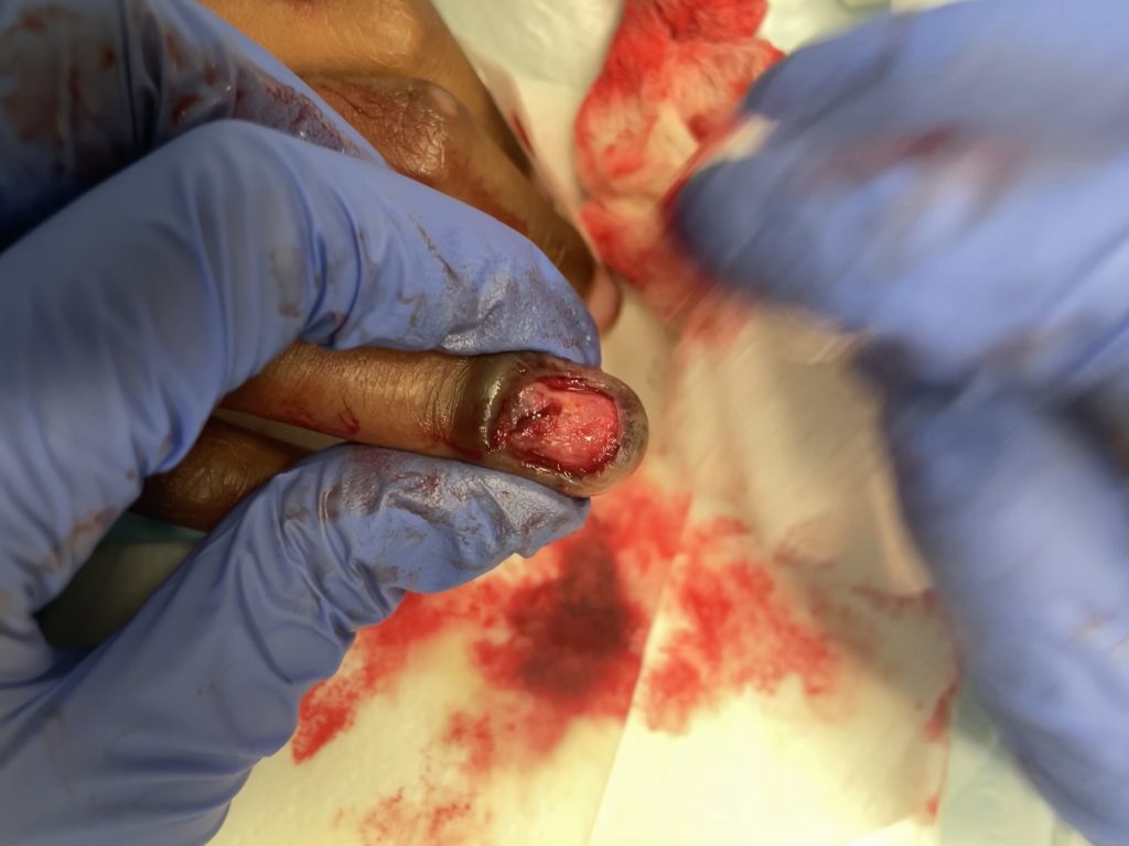
Figure 4: Visible nail bed laceration distal to the nail fold.

Nail bed laceration s/p repair

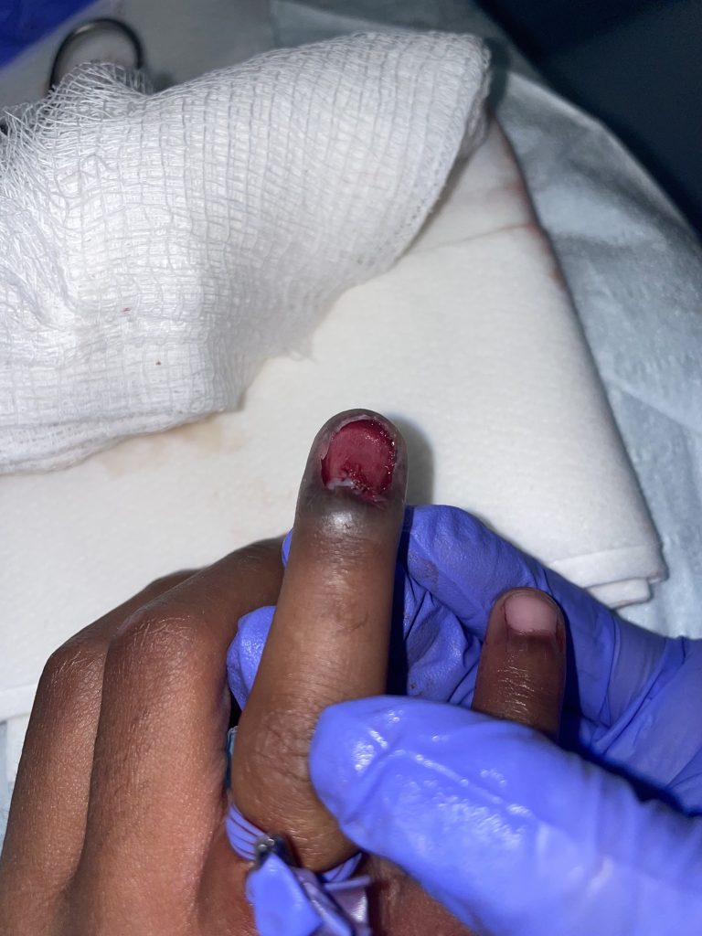
Figure 5: S/p nail removal and nail bed repair.

Discharge Instructions:

  • Keflex for 1 week
  • Keep dressing on for 2 days, followed by daily dressing changes with bacitracin/ xeroform/ kling wrap and splint.
  • Elevation of hand as much as possible
  • Follow-up PRS clinic in 1 week

Follow Up:

Patient seen in clinic 10-days post-injury with good wound healing. Education provided to both patient and mother that while the distal phalanx does not require operative fixation, it can be extremely tender and painful as it heals due to swelling and hypersensitivity. Also discussed that it may take up to 4-5 months for the nail to completely re-grow. Recommended using protective splint to minimize risk of re-injury, which can then be weaned over the course of the following six months.
Print Friendly, PDF & Email

References

Zook, Elvin G., Roxanne J. Guy, and Robert C. Russell. "A study of nail bed injuries: causes, treatment, and prognosis." The Journal of hand surgery 9.2 (1984): 247-252.
Germann, Günter, et al. "Fingertip and thumb tip wounds: changing algorithms for sensation, aesthetics, and function." The Journal of hand surgery 42.4 (2017): 274-284.
Martin-Playa, Patricia, and Anthony Foo. "Approach to fingertip injuries." Clinics in Plastic Surgery 46.3 (2019): 275-283.
https://pubmed.ncbi.nlm.nih.gov/6715836/
https://pubmed.ncbi.nlm.nih.gov/28372640/
https://pubmed.ncbi.nlm.nih.gov/31103072/

Related Articles