Pediatric Partial Thickness Hand Burn

Samantha Lund, MD
, Lauren Woldanski, MD
, Nicole Sommer, MD

History:

15-year-old male with no past medical history presented with burns sustained when he was lighting a bonfire with gasoline. He presented to an outside hospital via private vehicle where he was intubated for airway protection prior to transfer to the pediatric tertiary hospital.

Findings:

Integument of the bilateral upper extremities, face, trunk, and left foot was erythematous with scattered pale patches with intact blanching throughout. The volar surface of the arms were spared and burns were non-circumferential. Desquamation of the dorsal hand extended up the posterior arms to mid-humerus bilaterally with underlying moist dermis. Scattered areas of erythema over bilateral shoulders and right nipple. Left anterior lower extremity with erythema. Sensation unable to be tested due to patient being sedated and intubated. Intact capillary refill to the distal fingertips. Palpable radial and ulnar pulse.

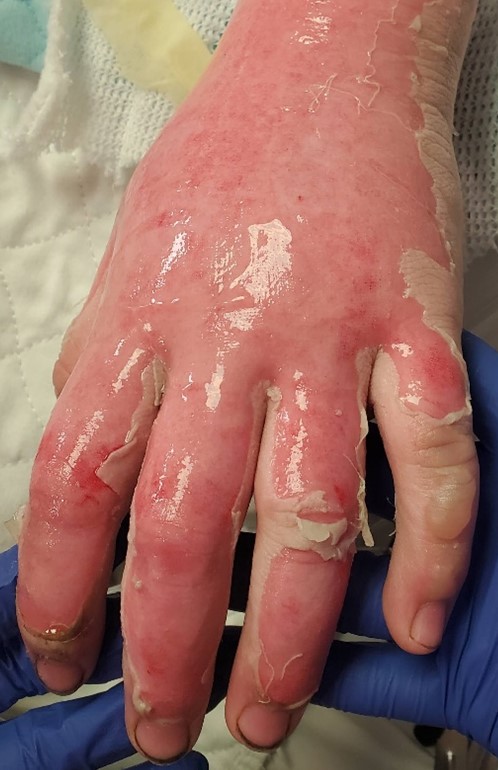
Fig.1. Left hand day of injury

Fig.2. Left upper extremity day of injury

Fig.3. Right upper extremity day of injury

Diagnosis:

16% TBSA (total body surface area) of mixed superficial and deep partial thickness burns to bilateral upper extremities, including the dorsum of bilateral hands, face, trunk, specifically the right nipple and shoulders, and the left foot.

Differential Diagnoses:

Partial vs full thickness burns

Workup Required:

Complete ACLS workup with airway assessment given mechanism with concern for inhalation injury and edema. Gold standard assessment for inhalation injury is flexible bronchoscopy. Accurate assessment of TBSA and burn depth is crucial in determining resuscitation and need for escharotomies and/or frequent neurovascular checks. Resuscitation is indicated in the pediatric patient if >15% TBSA. Pain control is a main component of a pediatric burn hospital stays and is usually multimodal with Tylenol, ibuprofen/Toradol, gabapentin, and opioids. Early enteral nutrition (if not requiring vasopressor support) is helpful to decrease needed volume of IV fluids to maintain adequate urine output (UOP). A foley is placed to measure hourly urine outputs in the first 24 hours. Management of hypermetabolic response with propranolol and oxandrolone is also often utilized.

Plan:

Resuscitation and burn care. If burns progressed to full thickness, would plan surgical excision with skin grafting (See Hand Burn, Deep). If not, would continue wound care until burns healed. Flexible bronchoscopy to assess for inhalation injury. Ophthalmologic evaluation given facial burns and concern for ocular involvement.

Expertise Needed:

Burn surgeon with hand experience

Treatment:

Burns are washed with warm, soapy water and blisters are unroofed. Desquamated skin removed. All wounds are assessed for blanching, turgor, texture, and moisture. Burns are dressed with bacitracin ointment, xeroform gauze, intersorb burn dressings, and wrapped in kerlix. Each digit individually wrapped in dressings on hands, with fluffs replacing intersorbs. The extremities are kept elevated as much as possible to reduce edema. Facial burns are managed with bacitracin (ophthalmic bacitracin near the eyes), to be applied prn to keep skin moist (typically 5-6 times throughout the day). Resuscitation with Parkland formula initiated. Accurate measurements of urinary output obtained every hour. Pain control with IV pain medication. Dressing changes can initially be done with conscious sedation in the pediatric ICU for pain control.

Daily monitoring of burns to evaluate for progression over initial 72 hours with daily dressing changes. Burns did not progress to full thickness, so surgery was not indicated. At this time, Mepilex Ag was used on all burned areas. The Mepilex was applied to each individual finger and secured in place with surginet. The Mepilex dressing can remain in place for 7 days due to the antimicrobial properties of the sliver. This reduces pain with dressing changes and provides an antimicrobial, moisture-wicking environment for wound healing.

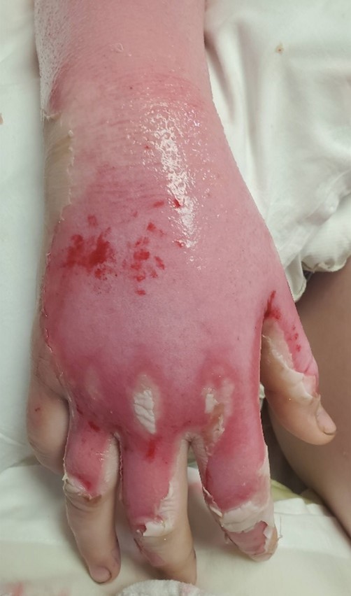
Fig.4. Right hand post-injury day 4

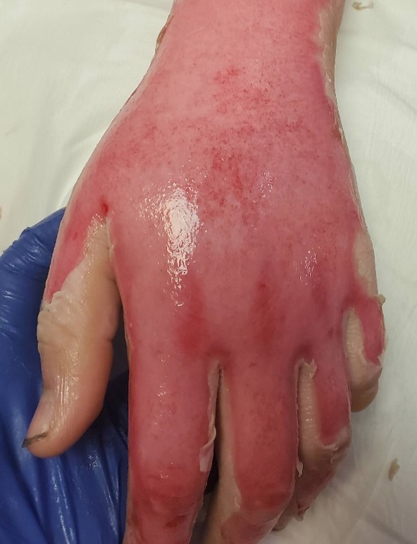
Fig.5. Left hand post-injury day 4

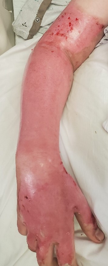
Fig.6. Right upper extremity post-injury day 5 and day of discharge

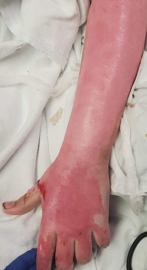
Fig.7. Left upper extremity post-injury day 5 and day of discharge

Follow Up:

The patient presented to clinic 1 week after discharge for Mepilex removal and assessment. At this time, the burns were entirely healed with reepithelization and no open wounds. It was recommended he apply lotion/Vaseline to the healed burns. Sun protection was discussed, as the new skin will be more sensitive and prone to burning.

He was seen 3 weeks after the injury for a final assessment.  At that time, sun protection and scar care were again discussed. There were no areas of hypertrophic scarring or hyperpigmentation. Discussed temperature sensitivity and skin fragility will resolve with time. He was discharged from clinic but instructed to follow up with any questions or concerns.

Print Friendly, PDF & Email

References

An open, parallel, randomized, comparative, multicenter investigation evaluating the efficacy and tolerability of Mepilex Ag versus silver sulfadiazine in the treatment of deep partial-thickness burn injuries
Handbook of Burns Volume 1
Antimicrobial dressings: Comparison of the ability of a panel of dressings to prevent biofilm formation by key burn wound pathogens
https://journals.lww.com/jtrauma/FullText/2015/05000/An_open,_parallel,_randomized,_comparative,.16.aspx?casa_token=VFwYKtfizBUAAAAA:J7MvmgbxrWefAGhyDTe3JnkSWtvsfGCM9O4cweaNM99ApOZpJ76szMvZn6v27dz8PmQtY-WIjpf5g1IQe4CBVzdu
https://link.springer.com/book/10.1007/978-3-7091-0348-7
https://www.sciencedirect.com/science/article/abs/pii/S0305417915001771

Related Articles