Open Foot Wound with Exposed Orthopedic Hardware

Aladdin H. Hassanein, MD, MMSc, FACS, Indiana University School of Medicine
, Connor J. Drake, BS; Indiana University School of Medicine

History:

51 year-old female with previous traumatic foot injury from motor vehicle collision. She had a previous open fracture of her right foot 1st metatarsal and underwent open reduction and internal fixation 2 months ago. She has a history of coronary disease with a coronary stent. She presented to an orthopedic surgery clinic with exposed hardware.
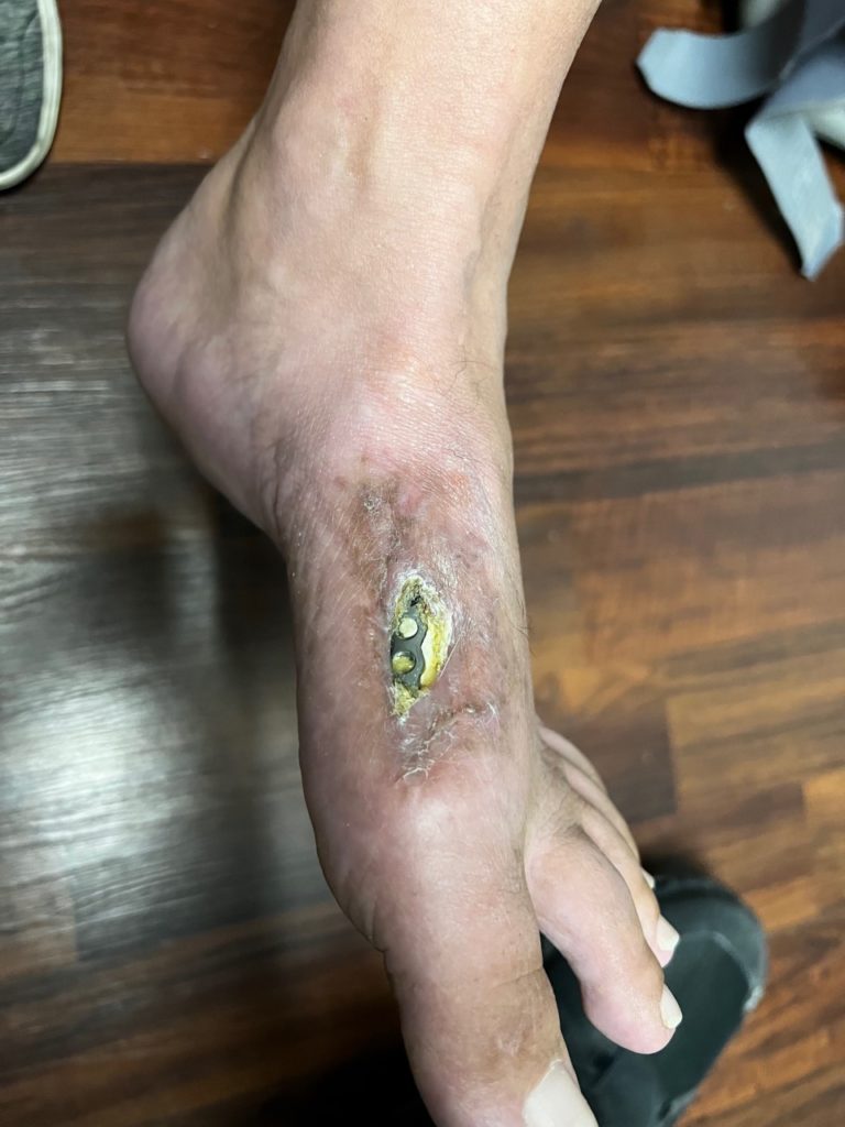
Fig.1.  Right dorsal foot wound over the 1st metatarsal with exposed hardware, tendon and bone.

Findings:

Exposed hardware right medial first metatarsal with no evidence of infection. There is hypertrophic scar surrounding the exposed hardware.

Diagnosis:

Right foot wound with exposed hardware over 1st metatarsal.

Differential Diagnoses:

Foot wound with exposed hardware; no evidence of contamination or osteomyelitis beyond the exposed area.

Workup:

Right foot x-rays exhibited nonunion of the first metatarsal fracture and evidence of osteomyelitis.

Plan:

Orthopedic surgeon  to remove exposed hardware, debride nonviable bone, and place antibiotic cement spacer. Soft tissue reconstruction requested. Options include 1) amputation, 2) free flap reconstruction, 3) local flap.

Expertise Needed:

A  foot wound along with a distal third of the leg wound with exposed bone/hardware often requires free tissue transfer reconstruction. However, in this patient who was not a good free flap candidates, a local flap appeared possible. The flap chosen was the bi-pedicled advancement flap. This was technically more simple and less complicated than a free flap.

Treatment:

Orthopedic surgeon performed a debridement, removal of the exposed hardware, excision of nonviable/osteomyelitic bone, and placement of antibiotic cement spacer.
The wound was assessed. A full thickness skin flap was raised lateral to the wound leaving dorsal tendons down with peritenon intact. Superficial dorsal foot veins were ligated as needed. Care was taken not to injury the skin flaps or over dissect the tissues. A counter incision was made to develop the bipedicle flap. This allowed for potentially a 2:1 length to width in the lower extremity as a random flap rather than 1:1 for reliability because of the dual blood supply. The skin flap was moved to the wound and a skin graft is placed at the donor site of the bipedicle flap so as to avoid opposing forces on the flap and minimize tension. Tension was avoided in order to minimize risk of flap necrosis. The flap was carefully inset tension free and a bolster placed on the skin grafted donor site.

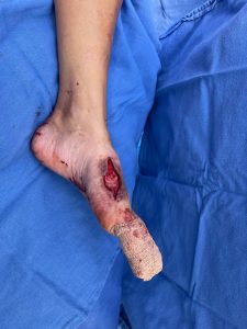
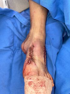
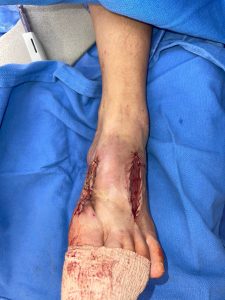
  Fig.2. Right dorsal foot wound. (Left) Following debridement and cement spacer placement, there was exposed antibiotic cement and bone. (Center) Bipedicled flap was raised as a skin flap adjacent to the wound. The dual dermal blood supply  improved reliability and allowed for an increased length to width ratio compared to a standard unipedicled random flap. (Right) The flap donor site was skin grafted to minimize flap tension.

Follow Up:

The bolster was removed from the skin grafted donor site 1 week later. The patient was scheduled for removal of the cement spacer and bone graft when the wound was completely healed.. The flap would need to be re-elevated to facilitate this.
Print Friendly, PDF & Email

References

Baker BG, Oudit D. The modified bipedicled flap for reconstruction of oncological skin defects of the trunk and extremities. J Plast Reconstr Aesthet Surg. 2020 May;73(5):913-920. doi: 10.1016/j.bjps.2019.11.030. Epub 2019 Nov 28. PMID: 31983663
Dujon DG, Khan UD, Aslam S. Bipedicle flaps: simple solutions for difficult problems in the extremities. Br J Plast Surg. 1997 Dec;50(8):641-5. doi: 10.1016/s0007-1226(97)90511-7. PMID: 9613408.
Granzow JW, Li A, Suliman A, Caton A, Goldberg M, Boyd JB. Bipedicled flaps in posttraumatic lower-extremity reconstruction. J Plast Reconstr Aesthet Surg. 2013 Oct;66(10):1415-20. doi: 10.1016/j.bjps.2013.05.019. Epub 2013 Jun 17. PMID: 23786879.
https://www.jprasurg.com/article/S1748-6815(19)30518-2/fulltext
https://www.jprasurg.com/article/S0007-1226(97)90511-7/fulltext
https://www.jprasurg.com/article/S1748-6815(13)00294-5/fulltext

Related Articles