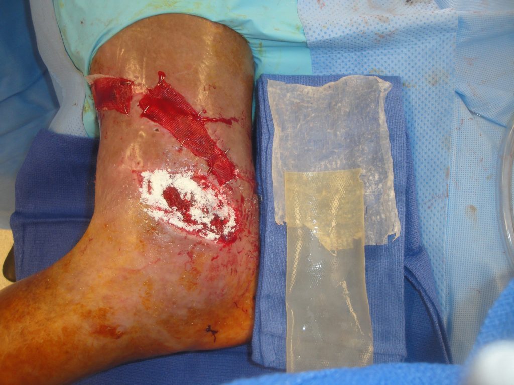
Open Ankle Wound with Exposed Orthopedic Hardware

Bruce A. Kraemer, M.D.
History:

This 80-year-old male weighing 475 pounds (BMI= 64) fell at home while getting out of his bathroom shower.  Due to his large size, Emergency Medical Services had to cut him out of his bathtub where he had been trapped for several hours.  He was hospitalized and cared for by the Orthopedic Service for 1 month prior to consulting the Plastic Surgery service for treating his open right ankle wound
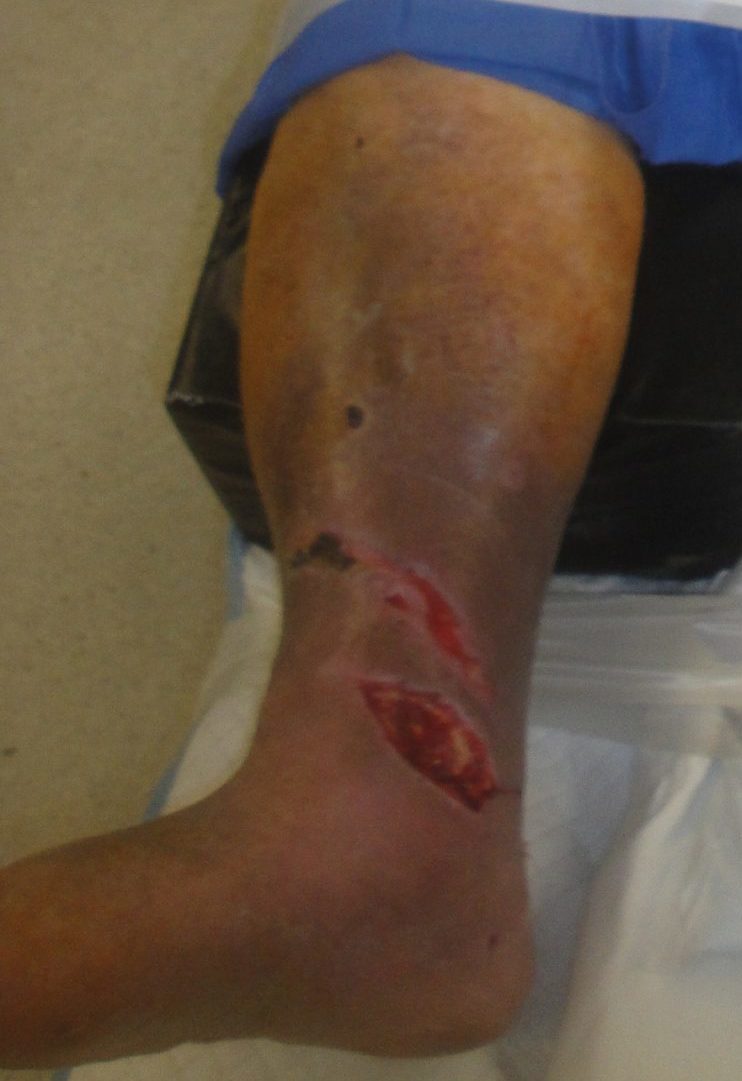
Fig.1. Patient with open right ankle wound

.Initial wound management with NPWT- note the hydrocolloid dressing was being used to protect the fragile venous stasis ulcer prone skinDuring this admission he had been treated twice for his acutely fractured ankle.  Initially it was managed with both internal and external ankle fixation but 1 week later it was converted to total internal fixation after he refractured it while getting up and trying to ambulate on his own.  His ankle wound had been managed with negative pressure wound therapy by the Orthopedic Surgical service prior to consultation

In addition to being an octogenarian, he had asthma, COPD, depression, anxiety, type 2 diabetes, hypertension, hyperparathyroidism, osteoporosis, psoriasis, marked venous stasis disease, an old Charcot foot injury of his left foot and he stopped smoking at age 73.  He has a family history of having a Protein S deficiency.
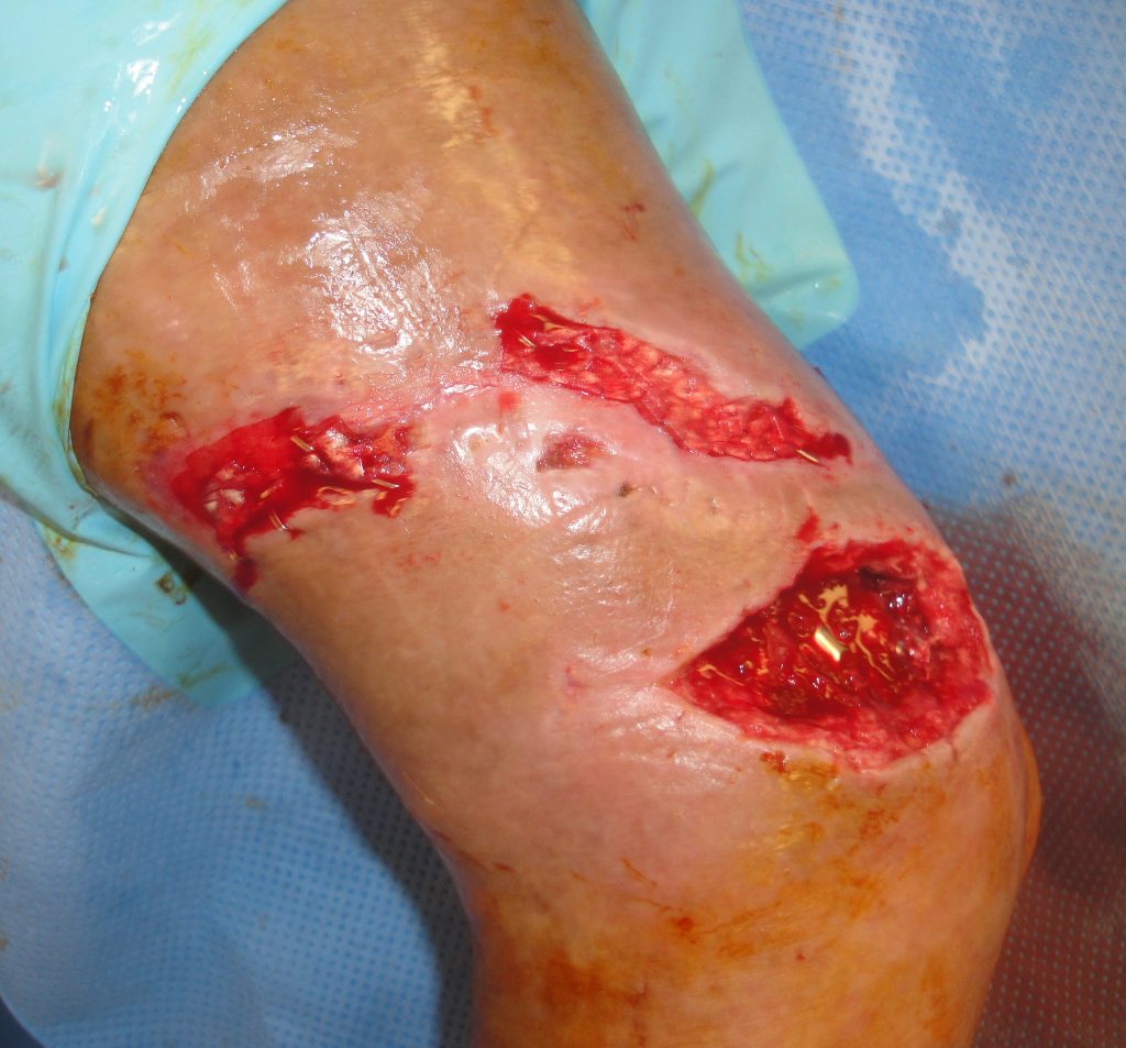
 

Fig.2. Open Ankle wound with exposed hardware plus proximal injuries
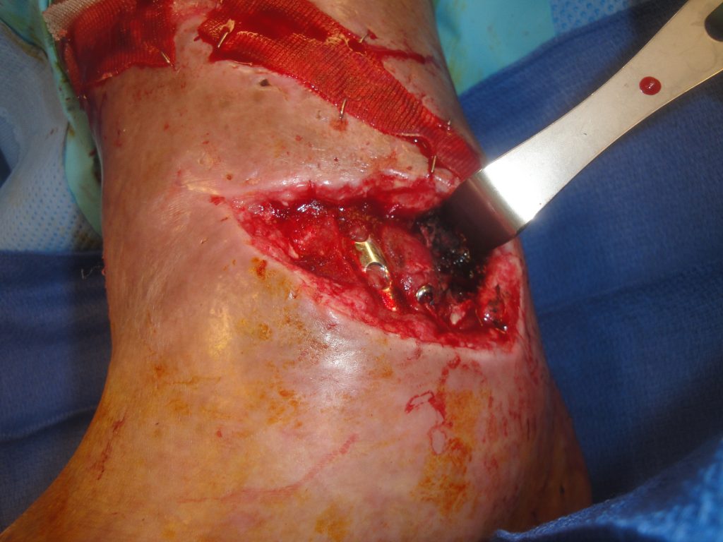
Fig.3. Open ankle wound with the degloving aspect demonstrated

Findings:

.The large full thickness medial right ankle wound was 12 cm. in length with exposed metal hardware, bone, tendons and had a degloving component extending around and down into the ankle joint.  There was a 17 cm long oblique wound above the open ankle wound

Recent wound cultures were positive for Enterobacter cloacae, Bacteriodes fragilis & Diptheroids

Diagnosis:

Significantly colonized open right medial ankle joint wound with exposed bone, tendon and hardware in an elderly morbidly obese medically debilitated patient.

Differential Diagnoses:

None- Subacute ankle wound injury needing definitive wound management.  Clinical judgement was that the wound was significantly colonized but not critically colonized with bacteria.

Fig.4. Initial Fracture Management

Fig.5. Reoperative Fracture Management

Workup Required:

The patient had been well managed by the Orthopedic service with serial x-rays, optimal fracture management and serial wound cultures which unfortunately were positive.  His host of medical problems were being optimized by the medical service but there was was no reversing his obesity and marked venous stasis disease.

Plan:

The treatment goal was to attempt limb salvage as he would most likely never have any chance at independence again if he was treated with a below-the-knee amputation given his associated right Charcot foot problem and obesity.  He was considered an an extremely poor microsurgical candidate and local and regional flaps also seemed out of the question.  Given the degree of tissue loss over his ankle wound, Extracellular Matrix (ECM) wound device use was decided upon as the preferred method of wound management

Expertise Needed:

A plastic surgeon with particular interest in soft tissue reconstruction should assess the patient.  Knowledge of the use of Extracellular Matrix Wound Healing Devices is desired as these devices facilitate healing through a process termed “site-specific constructive remodeling”, the formation of more normal tissue much like the tissue that was lost and being replaced.  This M-2 macrophage directed healing is highly desirous in treating a colonized wound with full thickness tissue loss.  These ECM devices, especially the Urinary Bladder Matrix-Extracellular Matrix (UBM-ECM) device which comes in several formulations and have different timelines of activity have been used by the author in a combined fashion to produce a sustained healing response in such a wound.

Treatment:

The medial right ankle wound was first cleansed and judiciously debrided with hydrosurgical dissection.  Next 500 mg of the UBM-ECM powder was placed into the open joint wound along with the10 x 15 lyophilized 2-layer UBM-ECM sheet so that the wound device was tucked under and around the open joint wound margins.  Next, a 6 x 15 cm 6-layer vacuum-pressed UBM-ECM sheet was cut into strips which were then layered over the open joint wound in multiple layers and over the upper wound as a single layer.  The 6-layer device as well as the retaining petroleum impregnated gauze were secured with staples prior to applying a hydroconductive sheet for a secondary dressing and an outer polyurethane sheet outer dressing.  Total operative time was 45 minutes.
The patient was continued on appropriate antibiotics for 6 weeks after UBM-ECM wound device placement.

Fig.6. Open ankle wound with the UBM-ECM device to be utilized in management- the powder is already in place in the wound
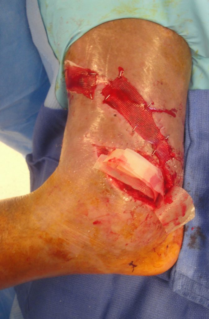
Fig.7. The lyophilized powder is now in the wound and multiple vacuum pressed sheets are to be stapled over this
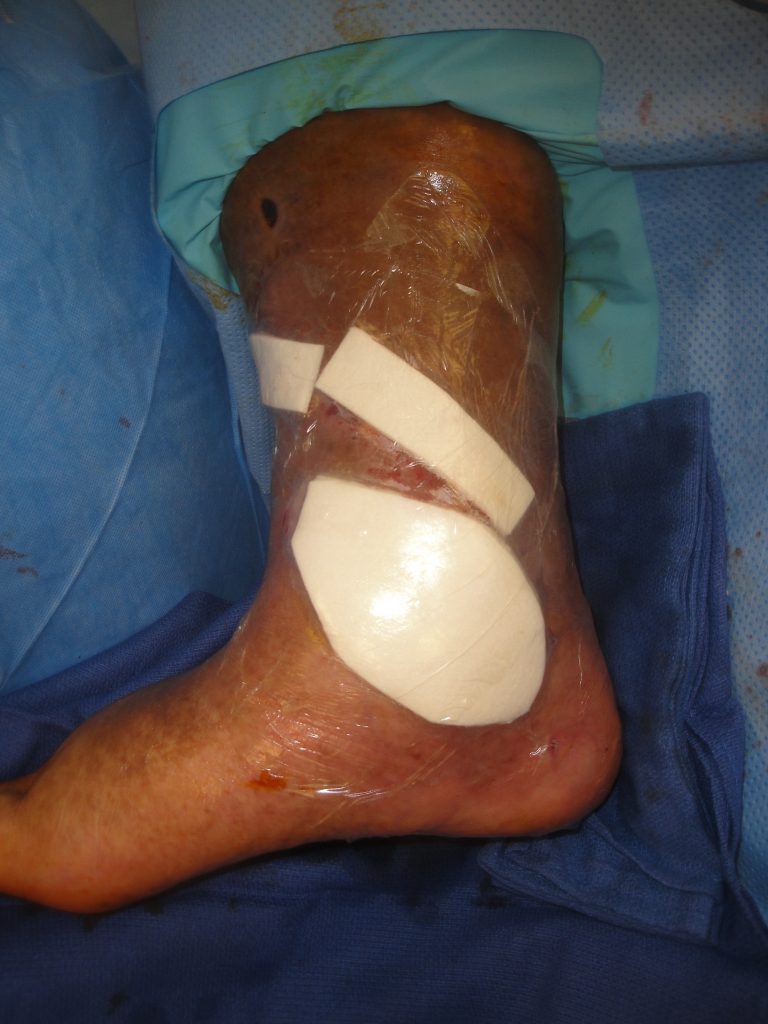
Fig.8. Secondary dressings in place to stent the UBM-ECM into the wounds.

Follow Up:
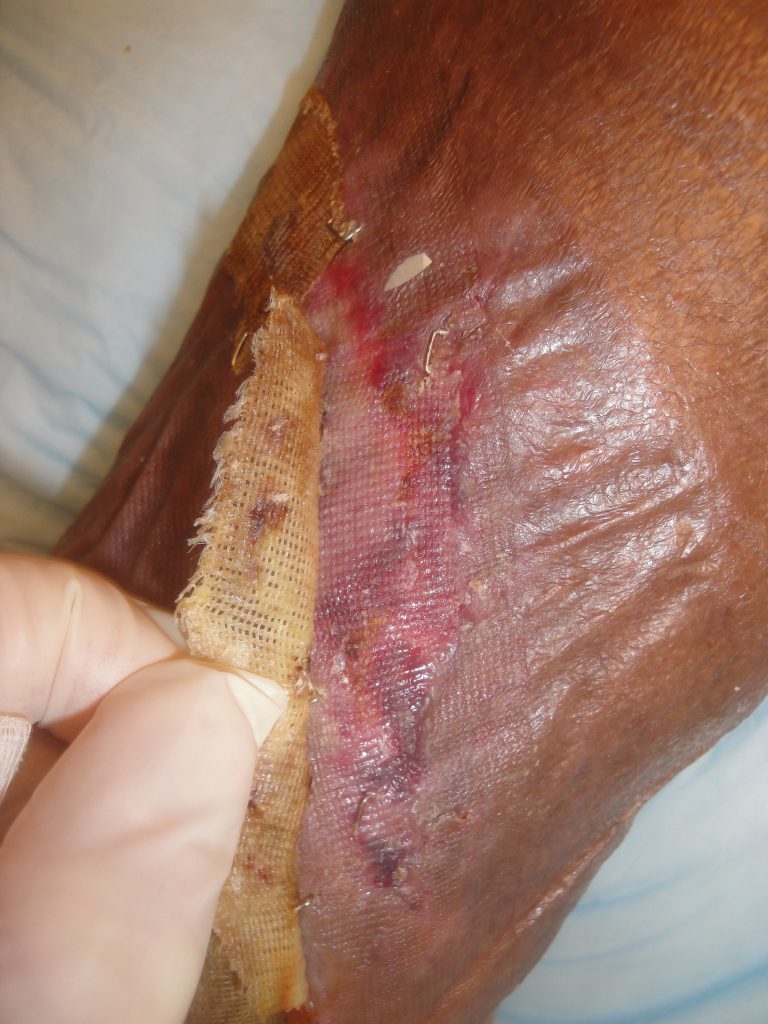
The patient had the polyurethane sheet dressing reinforced as needed for the first 3 weeks  At that time the wound appeared to be very dry as seen in Figure ___.  The topical wound care was then changed to daily hydrogel with a non-adherent secondary dressing.  His wound was healed at 3 months and final follow-up photos were taken at 4 months.  Overall care involved minimal follow-up care and no intensive surgical intervention or prolonged operative procedures.

Fig.9. Upper wound healing at 3 weeks- Healed
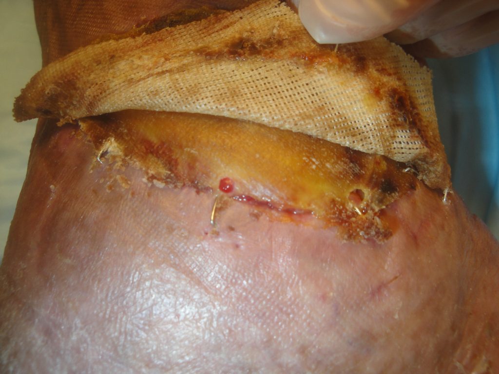
Fig.10. Open ankle wound healing at 3 weeks- UBM-ECM device adherent to the wound margin but overall too dry
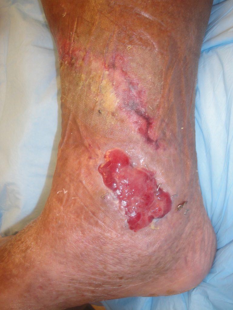
Fig.11. Right medial ankle wound 1 month later after daily hydrogel application
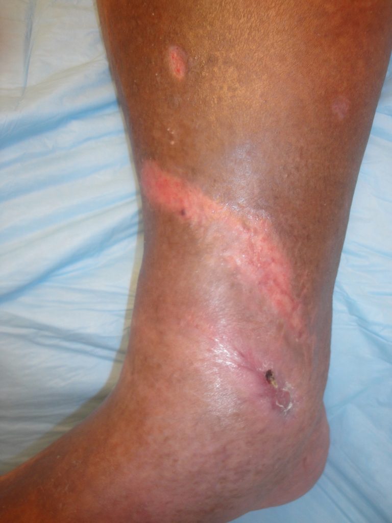
Fig.12. Right ankle wound appearance 4 months after UBM-ECM treatment- Total Healing with minimal scarring.

Print Friendly, PDF & Email

References

Geiger SE, Deigni OA, Watson JT, Kraemer BA. Management of Open Distal Lower Extremity Wounds With Exposed Tendons Using Porcine Urinary Bladder Matrix. Wounds- A Compendium of Clinical Research & Practice 2016;28(9):308-316.
Kraemer BA, Geiger SE, Deigni OA, Watson JT. Management of Open Distal Lower Extremity Wounds with Concomitant Fracture Using Porcine Urinary Bladder Matrix. Wounds- A Compendium of Clinical Research & Practice 2016; 28(11):387-394.
Kraemer BA: Management of Complex Distal Lower Extremity Wounds Using a Porcine Urinary Bladder Matrix (UBM-ECM). In: Shiffman MA, Low M Eds. Plastic and Thoracic Surgery, Orthopedics and Ophthalmology. Cham, Switzerland: Springer International Publishing AG; 2020: 3-29.
https://pubmed.ncbi.nlm.nih.gov/27701126/
https://pubmed.ncbi.nlm.nih.gov/27861131/
https://link.springer.com/book/10.1007/978-3-030-10710-9

Related Articles