Findings:
1.8×1.5cm full thickness wound left nasal tip down to nasal mucosa centrally and tapering on the sides.
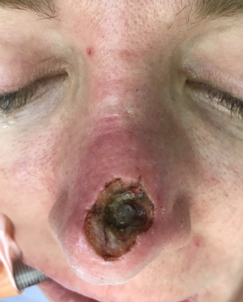
FIgure 1. Nasal tip defect.
Treatment:
The patient did not want a forehead flap, thus an extended local V-Y flap was chosen. The nose and incision lines were injected with local anesthesia with epinephrine. She was then prepped and draped in an aseptic manner. The wound edges were debrided with a 15-blade and iris scissors. The flap was incised and then was mobilized along its course. The distal part of the flap was completely elevated from the underlying deep structures to allow for complete mobilization. Care was taken to preserve perforators along the nasolabial region. The flap was inset with 4-0 monocryl deep dermal and a combination of simple interrupted and running 5-0 prolene sutures covered with steri strips. At the end of the procedure, the flap was well perfused.
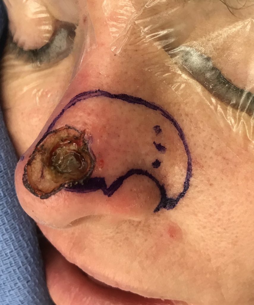
Flap markings. The length of the flap measures 1.5-2 times the longitudinal diameter of the defect. The long axis is place in the direction of maximal mobility. The flap width is greater than the width of the defect due to the extension component of the flap. The length of the extension component is the width of the defect such that the extension limb hinges down as a transposition flap, closing the distal-most portion of the defect.
Raising the flap. Note the angular artery perforators at the base of the flap.
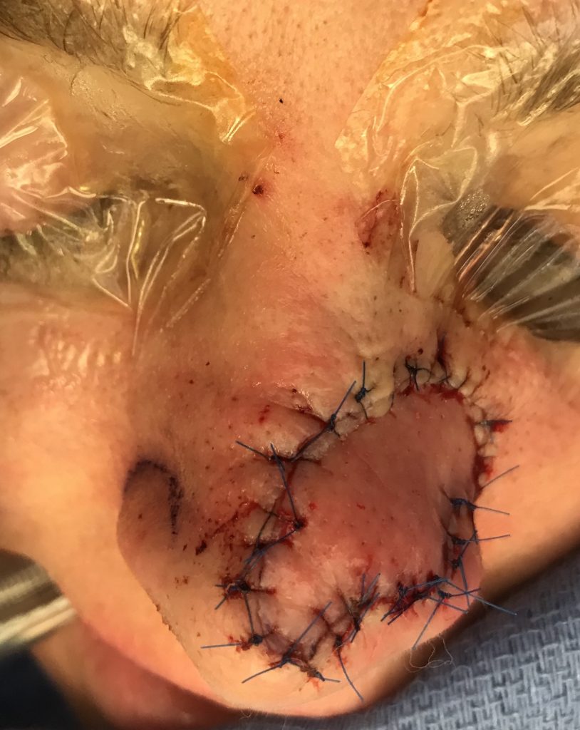
Flap inset.
Follow Up:
She healed uneventfully and sutures were removed at her one week postop visit. At 1 month postop her incisions were well healed. She had minimal amount of distal “pin-cushioning” of the flap and some asymmetry between the two nostrils but her nasal valve was patent. Overall, she was pleased with her result and plans to continue with scar massage.
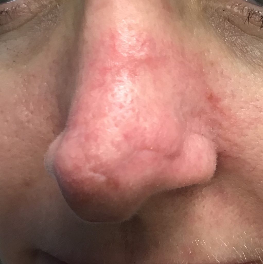
1 month Post op – anterior view
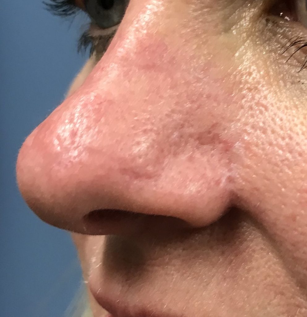
1 month Post op – lateral view
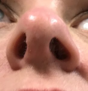
1 month Post op – worm’s-eye view
