Multiple Fingertip Amputations

Bruce A. Kraemer, M.D.
History:
53-year-old truck driver who sustained transverse amputations of his right index, long and ring fingertips by the fan-belt of his truck engine.  He is right-handed, a non-smoker and had no prior hand trauma.  In addition to his truck driving duties, he must be able to load and unload the trucks he drives.

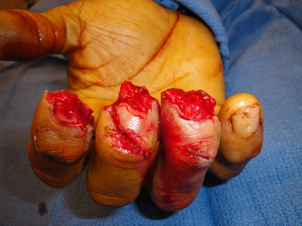
Fig.1. Right Index. Long, and Ring Fingertip injuries at the time of injury

Findings:

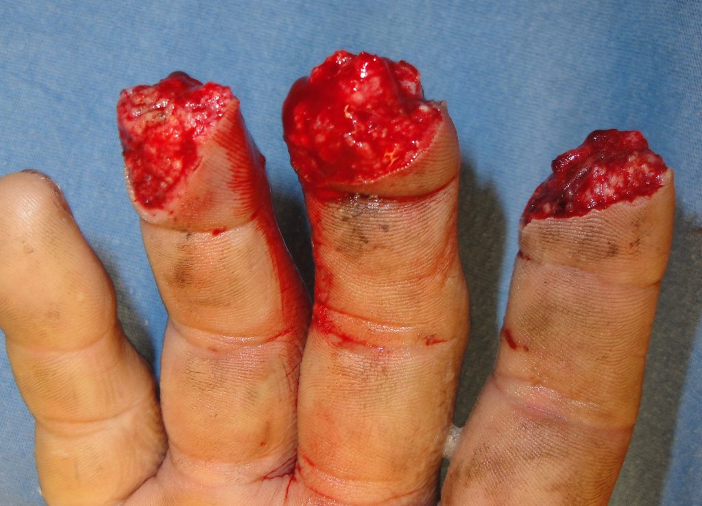
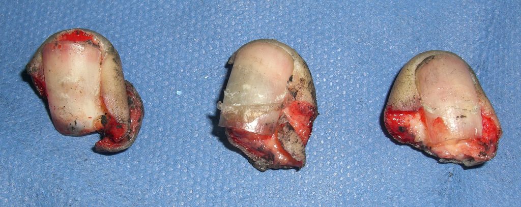
Fig.2. Avulsion fractures of the distal phalanx of the right index, long, and ring finger tips with injuries to the injured parts precluding microvascular reattachment.
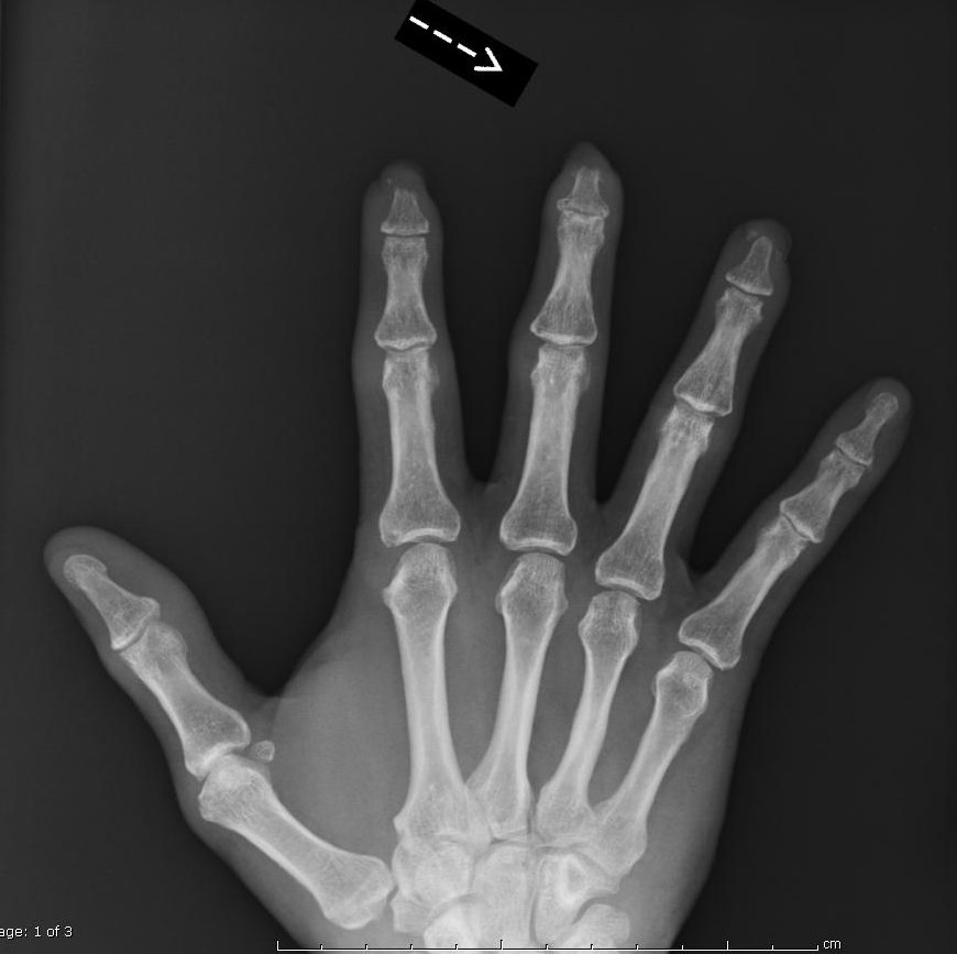
Diagnosis:

Allen Type IV finger fingertip amputations of the right index, long and ring fingertips.

Differential Diagnoses:

None- Acute Injury

Workup Required:

Fig.3. X-rays of the right hand to evaluate proximal injuries and possible retained foreign bodies

Plan:

The goal was to preserve maximal digital length with stable, sensate, durable healing.  The day prior, the author had been shown the UBM-ECM topical wound powder which in 2010 was being recommended for fingertip amputations- 20 mg. placed on alternate days until wound closure.  The patient opted to try this treatment not knowing how long it would take to heal his fingers but he did not want any shortening.

Expertise Needed:

As fingertip injuries are amongst the most common hand injuries, these injuries are most often treated by a hand surgeon.  Newer topical healing methods such as use of Extracellular Matrix wound care dressings (e.g.- UBM-ECM) is desirous as they promote M-2 vs. standard M-1 macrophage directed wound healing.

Treatment:

Alternate day application of 20 mg. UBM-ECM powder was applied to each fingertip with hydrogel applied on top of a petroleum impregnated gauze to maintain a moist environment.

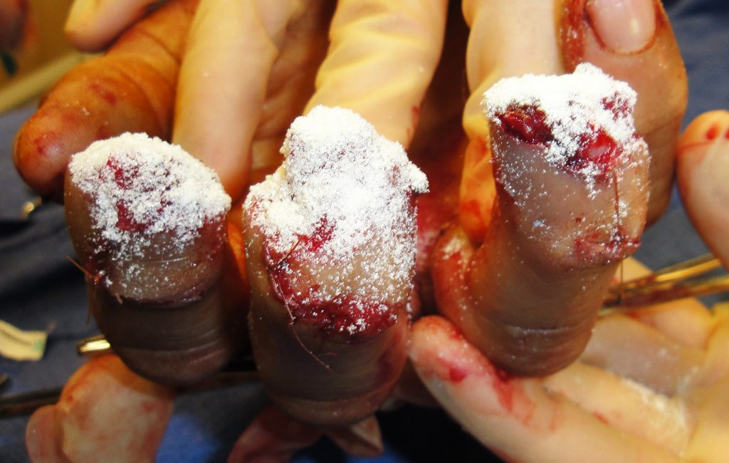
Fig.4. Fingertip appearance at the time of first application of UBM-ECM powder

Follow Up:

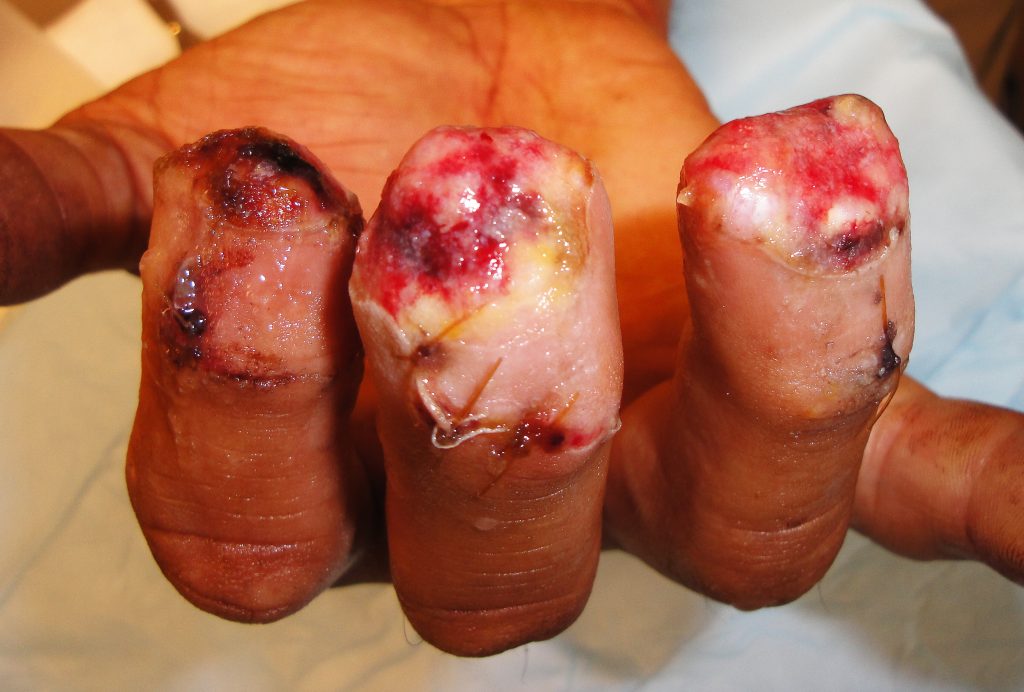
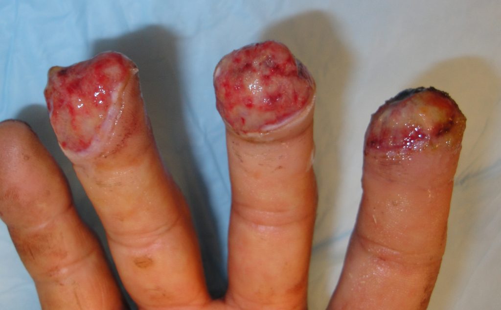
Fig.5. Fingertip appearance after the first week and 3 treatments

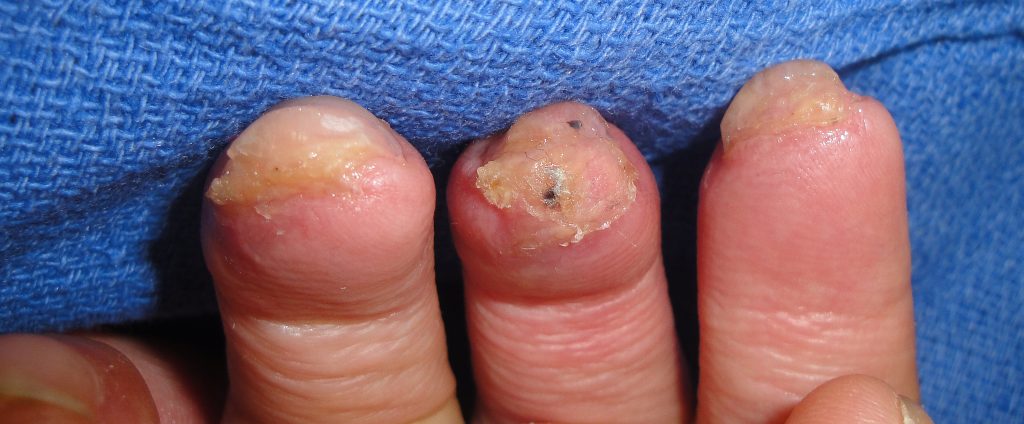
Fig.6. Fingertip healing at 2.5 months.

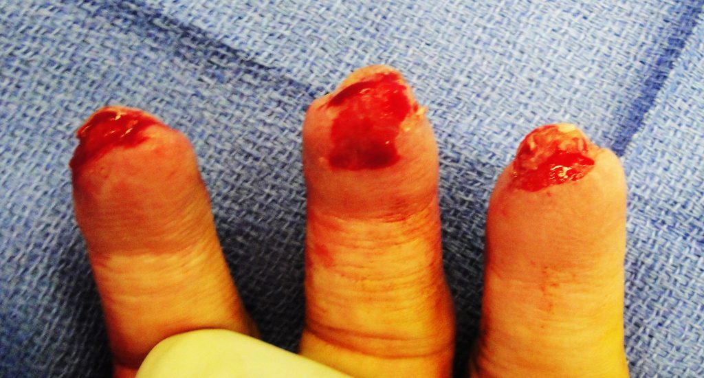
Fig.7. Fingertip appearance after debriding the thickened distal tissues and opening up the distal tips for placement of additional UBM-ECM powder.

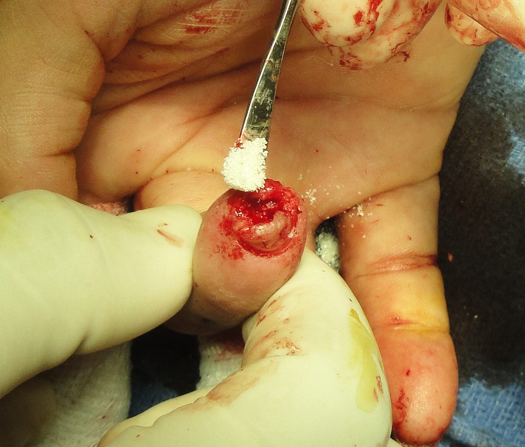
fig.8. After opening the distal fingertip- additional powder was placed under the fingernail and over the distal bone.

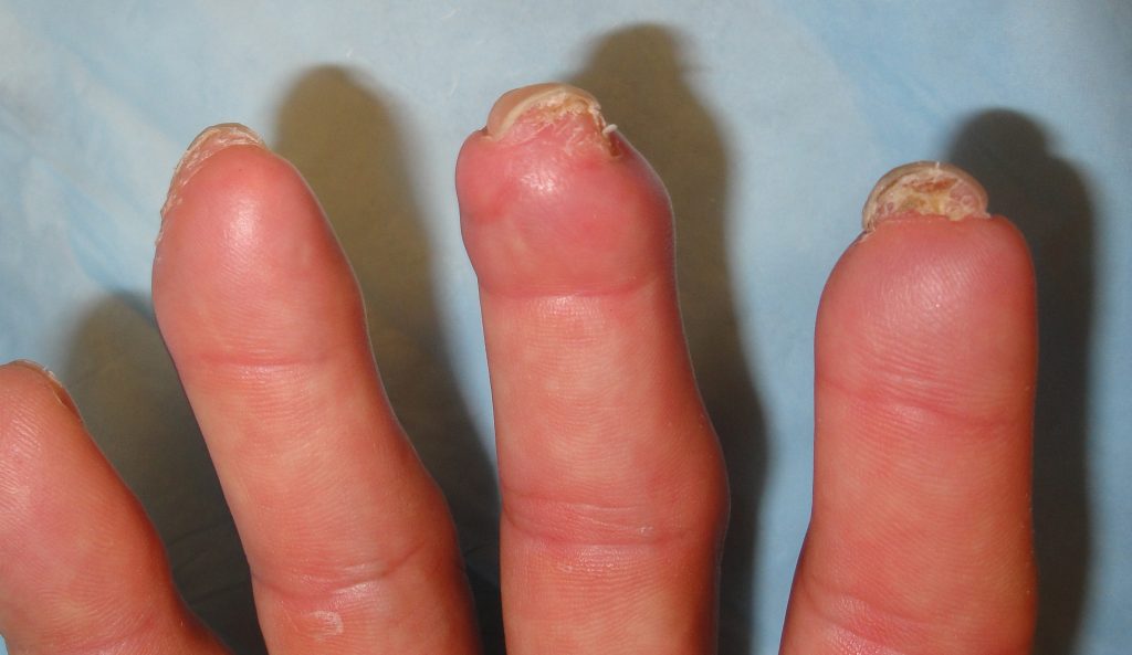
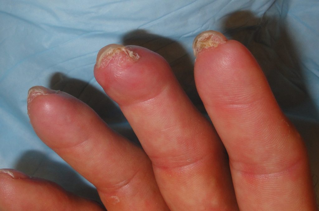
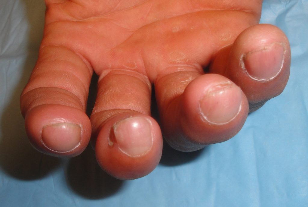
Fig.9. Fingertip Healing at 6 months post injury

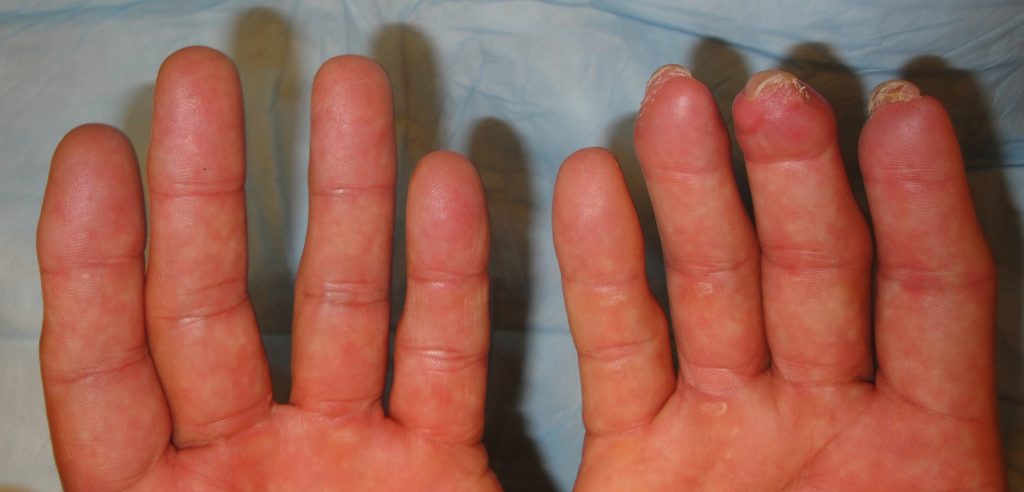
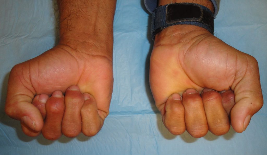
Fig.10. Fingertip appearance and comparison at 1 year post-injury.

The patient had stable healing and was able to resume his prior employment after 4 months.

Print Friendly, PDF & Email

References

Geiger SE, Deigni OA, Watson JT, Kraemer BA. Management of Open Distal Lower Extremity Wounds With Exposed Tendons Using Porcine Urinary Bladder Matrix. Wounds- A Compendium of Clinical Research & Practice 2016;28(9):308-316.
Kraemer BA, Geiger SE, Deigni OA, Watson JT. Management of Open Distal Lower Extremity Wounds with Concomitant Fracture Using Porcine Urinary Bladder Matrix. Wounds- A Compendium of Clinical Research & Practice 2016; 28(11):387-394.
Behrens J, Kraemer BA. Abstract: Urinary Bladder Matrix-Extracellular Matrix (UBM-ECM) for Management of Complex Upper Extremity Wounds. Plast Reconstr Surg Global Open 2018;6(9 Suppl):51.
https://pubmed.ncbi.nlm.nih.gov/27701126/
https://pubmed.ncbi.nlm.nih.gov/27861131/
https://www.ncbi.nlm.nih.gov/pmc/articles/PMC6212145/

Related Articles