Multiple Complex Degloved Finger Wounds

Bruce A. Kraemer, M.D.

History:

An otherwise healthy 24-year-old male sustained a severe injury to the dorsum of his right hand fingers after the truck he was driving rolled off a bridge.  He had a congenital left elbow dislocation so his right hand was truly his dominant hand.  He was a non-smoker, had no other significant medical problems and did not take any routine medications

Findings:

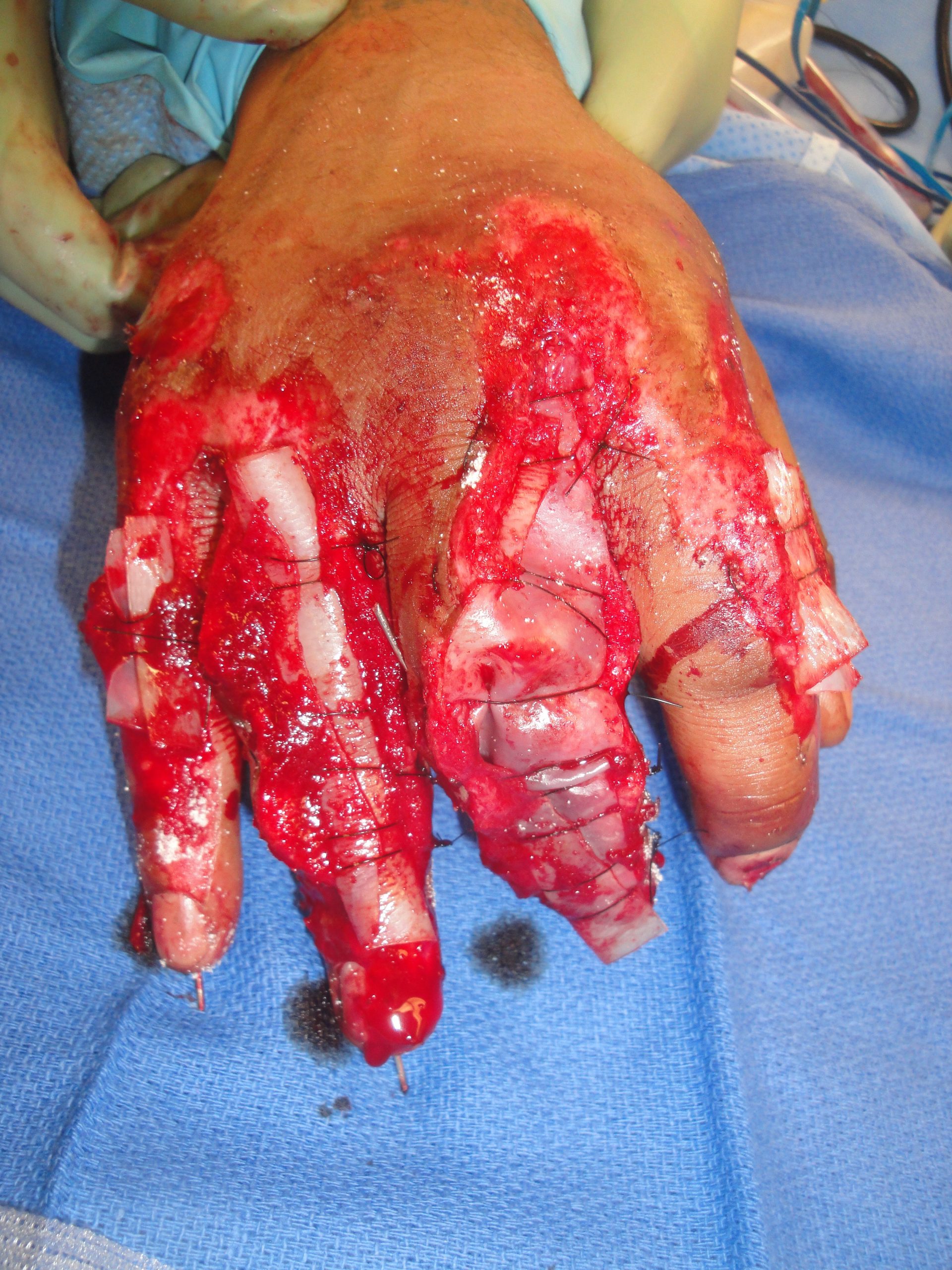
There were open wounds of all 4 fingers centered over the dorsum of the proximal interphalangeal joints with the most severe injuries to the long and ring fingers where the tissues overlying the PIP joints was missing

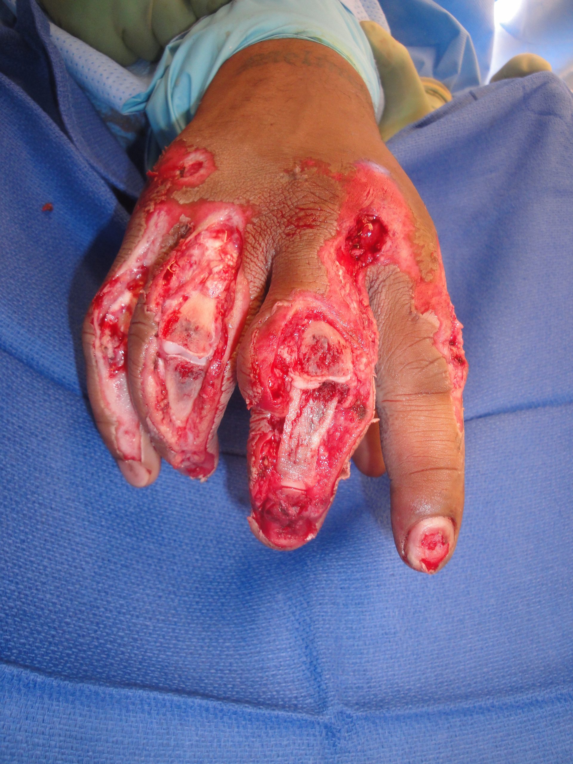
Fig.1. Intraoperative view of the dorsal finger injuries on the day of injury

Diagnosis:

Multiple complex dorsal finger wounds involving skin, extensor tendons, bones, joints, and nailbeds plus unstable long, ring and little finger proximal interphalangeal joints.

Differential Diagnoses:

None- Acute Injury

Workup Required:

After sustaining a major roll-over vehicular trauma, the patient must be hemodynamically stabilized, and assessed for occult internal organ injury.  It is best to obtain clearance from a general surgical trauma service standpoint before proceeding with wound specific care.

Wound/Injury Specific assessment- The neurovascular status of the injured digits needs to be assessed and ultimate tissue viability determined.  With complex wounds such as these, it is best done in the operating room.  High quality PA, lateral and oblique x-rays of the digits, hand and wrist done preoperatively or in the operating room are essential to determine the degree of skeletal injury (fractures, joint malalignment or dislocation) and overall stability.  One should not only assess the obvious fractures but look for occult fractures of the digits, hand, and wrist.  One should also look for retained glass and metallic foreign bodies which could cause delayed healing.

Plan:

A bulky compressive dressing is applied to the injured hand to both splint it and control the bleeding while the patient’s potential other injuries in his rollover accident.  Rapid assessment of serious hand injuries is best done in the operating theater under regional or general anesthesia.  Placement of catheters for regional anesthesia allows for optimal operative as well as post-operative pain management and possible administration of vasodilating agents if needed.  The operative decision of tissue salvage versus completion amputation is best done by the most senior hand surgeon.

Complex multi-finger wounds may be treated with tissue options such as a variety of regional pedicled trunk flaps after syndactylization of the digits with subsequent debulking and separation in a series of operations over many months.  Single finger wounds could possibly be managed by local pedicled dorsal hand flaps, free fascial tissue flaps or dorsal foot flaps but less optimal for treating this patient.


Expertise Needed:

A hand surgeon with particular interest in soft tissue reconstruction should assess the patient.  Knowledge of the use of Extracellular Matrix Wound Healing Devices is desired as these devices facilitate healing through a process termed “site-specific constructive remodeling”- promotion of the formation of more normal tissue which often minimizes scarring.  This M-2 macrophage directed healing is highly desirous in treating a seriously injured hand.

Treatment:

Assessing tissue viability and cleansing of the wound bed with sharp surgical debridement coupled with pulsatile irrigation are essential first steps in management.  The remaining tissues were found to be viable and cleansed of all gross contamination. At this point the decision of complex free tissue flap reconstruction vs. tissue salvage with reconstruction vs. completion amputation proximal to the injured joints was considered.  The decision was made to reconstruct the fingers with the Urinary Bladder Matrix Extracellular Matrix (UBM-ECM) Wound Healing Device- MatriStem.

Treatment priorities were then as follows: 1) The PIP joints of the long, ring and little fingers required stabilization so K-wire pins were axially placed distal to proximal.  2) Open wound coverage was next. At the time this patient was treated, the only UBM-ECM formulations available were the powder and the vacuum-pressed multilayer sheets. A total of 1,000 mg of the MicroMatrix® UBM-ECM powder was used over the 4 fingers with a majority placed on the long and ring fingers.  Next a 6 x 15 cm UBM-ECM 6-layer vacuum pressed sheet was cut up and rolled up on itself and secured over the powdered open wounds with 3-0 nylon sutures in a bolster-type manner as seen in figures 2 & 3).  3) The UBM-ECM required a very moist environment and immobilization for optimal healing, so the fingers were wrapped with several layers of a Petroleum impregnated gauze with a piece of IV extension tubing secured over each finger dressing and then a larger bulkier gauze dressing.  Total operative time was 1 hour.

Longer Term Care

Saline (5 cc.) was placed into the IV tubing of each finger daily.  A dressing change under anesthesia down to the level of the petroleum gauze was done at post-injury day 5.  As good healing was demonstrated, daily generous applications of hydrogel as done along with hand elevation.

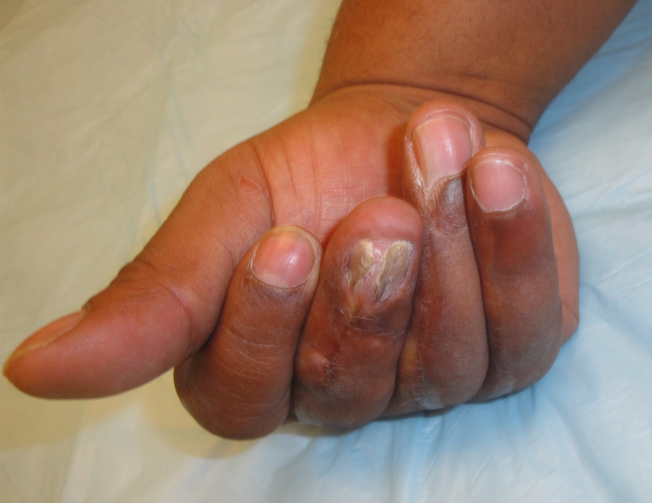
Exuberant healing was noted at various points which was treated with topical application of silver nitrate to lower the mounding granulation tissue down to the skin level which then allows and facilitates re-epithelialization of the wound.

UBM-ECM placement on finger wounds at the time of surgery

Follow Up:

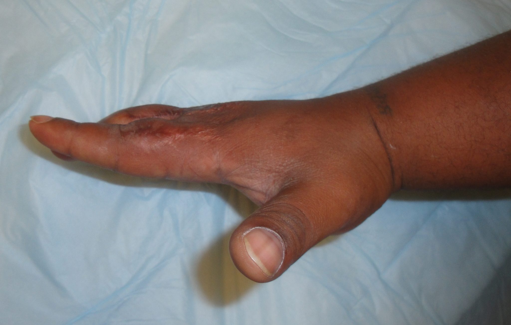
The hand wounds were all healed at 2.5 months and he worked at therapy to regain hand function.  He can extend his fingers, make a fist, and use his hand for writing

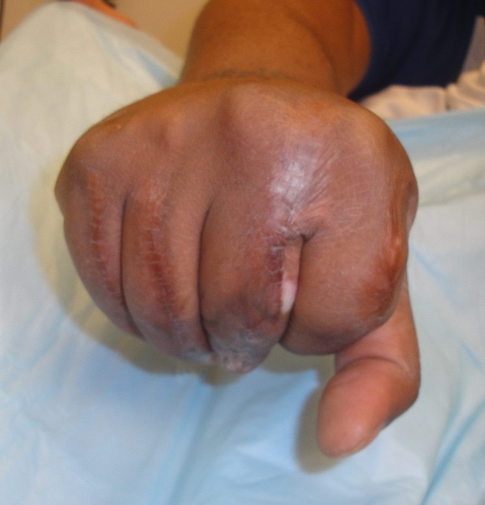
Fig.2. Finger wounds healing at 2 weeks

Fig.3. The wounds were kept moist with hydrogels as they initially developed exuberant granulation tissue which required several silver nitrate applications before final healing was achieved.

Clinical Note- This was the first time the device had been used by this author for treating such a complex multi-digit wound.  As amputation of both his long and ring fingers was seriously considered, this approach seemed reasonable at the time (July 2013).  Based on this case experience, the devices were used on more complex tissue reconstructions.  Of particular note was there was no additional donor site, excellent color match of the soft supple mobile tissues formed over the PIP joints and extensor tendon function seemed to be restored.

Retrospective note– While the appearance of the nail and nailbed was not really a significant initial treatment priority (and really isn’t a concern for the patient even today), had more time been given to placing more of both wound devices to the nailbed region.

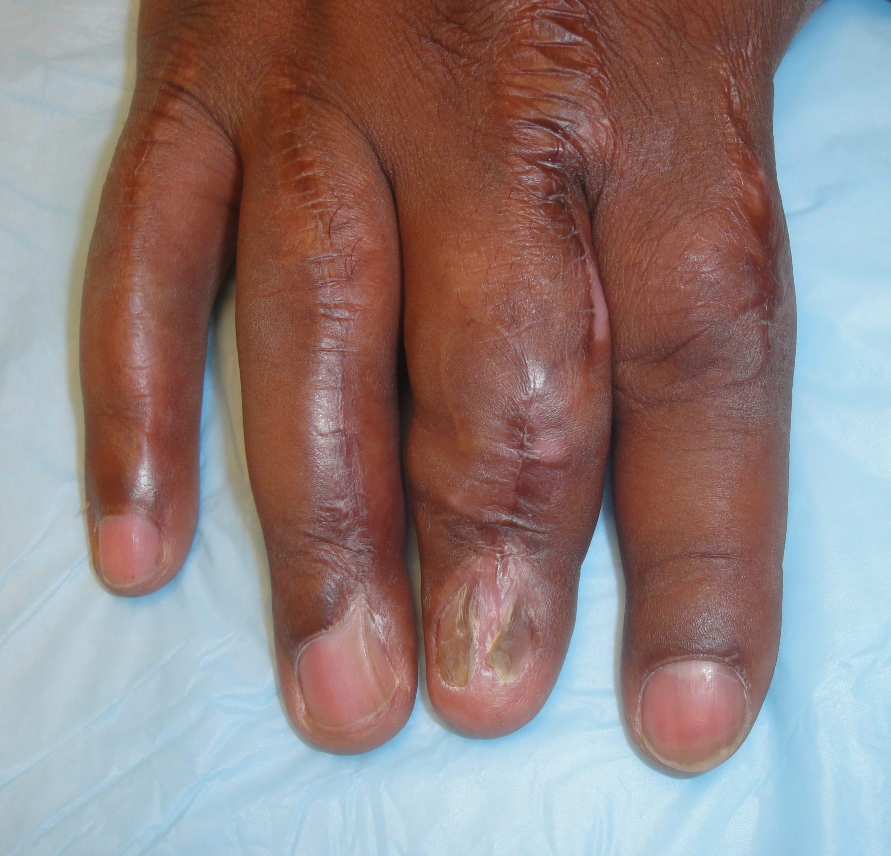
Fig.4.Final appearance of hand at 7 months (Wounds healed at 3 months)

Print Friendly, PDF & Email

References

Geiger SE, Deigni OA, Watson JT, Kraemer BA. Management of Open Distal Lower Extremity Wounds With Exposed Tendons Using Porcine Urinary Bladder Matrix. Wounds- A Compendium of Clinical Research & Practice 2016;28(9):308-316.
Kraemer BA, Geiger SE, Deigni OA, Watson JT. Management of Open Distal Lower Extremity Wounds with Concomitant Fracture Using Porcine Urinary Bladder Matrix. Wounds- A Compendium of Clinical Research & Practice 2016; 28(11):387-394.
Behrens J, Kraemer BA. Abstract: Urinary Bladder Matrix-Extracellular Matrix (UBM-ECM) for Management of Complex Upper Extremity Wounds. Plast Reconstr Surg Global Open 2018;6(9 Suppl):51.
https://pubmed.ncbi.nlm.nih.gov/27701126/
https://pubmed.ncbi.nlm.nih.gov/27861131/
https://www.ncbi.nlm.nih.gov/pmc/articles/PMC6212145/

Related Articles