Findings:
- Thumb: Fully intact
- IF (Index Finger): Missing soft tissue and nail over dorsal surface of distal phalanx. FDS/FDP intact.
- LF (Long Finger): Distal phalanx and associated soft tissue structures attached to proximal portion of finger with thin tissue bridge. The nearly amputated distal phalanx cold and not well-perfused. No dopplerable signals distally. O2 sat probe with 87% with poor waveform. FDS intact.
- RF (Ring Finger): Complete amputation through mid-shaft of fourth middle phalanx. FDS intact.
- SF (Small Finger): Missing soft tissue radially and volarly. Nail fully intact. FDS, FDP intact.
Median/radial and ulnar motor function grossly intact with thumb and finger adduction/abduction. Sensation intact in radial, median and ulnar distribution.
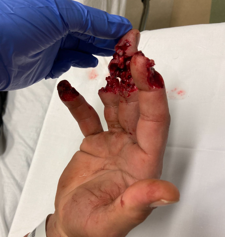
Figure 1: Day of presentation, extensive soft tissue and bony injury to ulnar four digits.
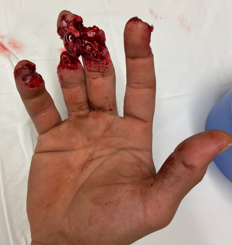
Figure 2: Day of presentation, extensive soft tissue and bony injury to ulnar four digits.
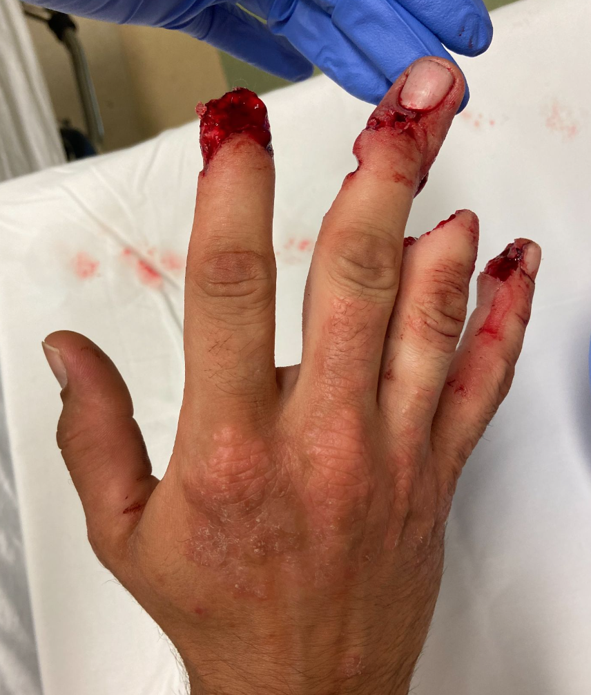
Figure 3: Day of presentation, extensive soft tissue and bony injury to ulnar four digits.
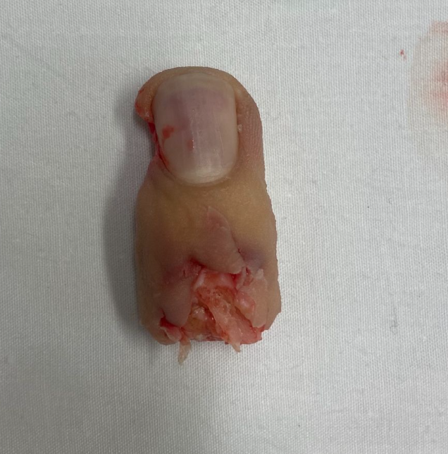
Figure 4: Amputated ring finger.
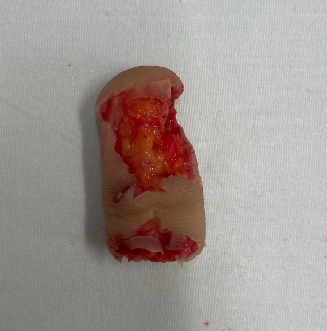
Figure 5: Amputated ring finger.
Workup Required:
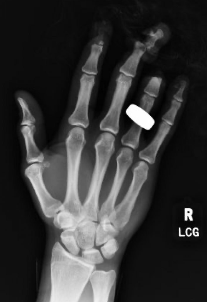
Figure 6: Hand XR (AP) on the day of injury.
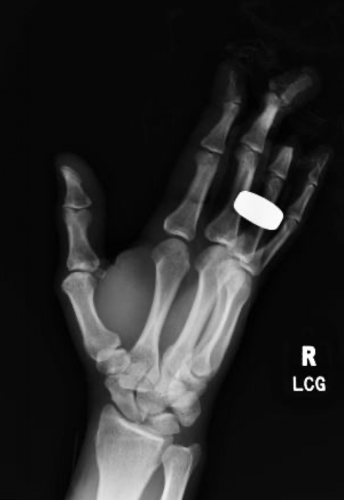
Figure 7: Hand XR (Oblique) on the day of injury.
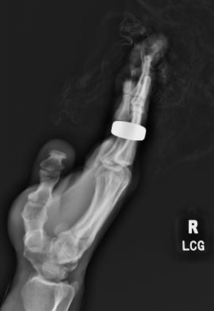
Figure 8: Hand XR (Lateral) on the day of injury.
- IF: Fracture at the base of distal phalanx
- LF: Comminuted fracture of third distal phalanx, distal phalanx being detatched at the level of DIPJ
- RF: Transverse amputation through mid-shaft level of middle phalanx
Treatment:
Digital block achieved by instilling 20cc of 1:1 mix of 2% lidocaine and bupivacaine to the affected four digits. Wound thoroughly irrigated with 500cc NS mixed with Betadine.
- Rongeur was used to shorten remaining RF middle phalanx to allow for enough soft tissue coverage. Skin closed with 4-0 chromic in simple interrupted fashion.
- Amputation was completed for LF while preserving as much soft tissue and skin to allow for coverage for middle phalanx. Skin flap was transposed over bone and was secured into place with 5-0 Monocryl. Full-thickness skin graft was harvested from the finger tip of the amputated LF. The graft was de-fatted with scissors as much as possible and was secured into place over proximal portion of remaining soft tissue. Skin graft and flap secured to place with 4-0 chromic.
All digits were again thoroughly irrigated again with 500cc NS. Patted dry.
- Occlusive dressing applied to IF and SF with bacitracin, Tegaderm, gauze, Alumafoam splint in DIPJ extension, all secured in Coban.
- LF and RF dressed with bacitracin, Xeroform, guaze and Coban.
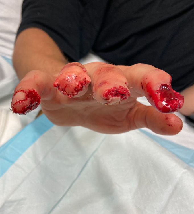
Figure 9: S/p repair.
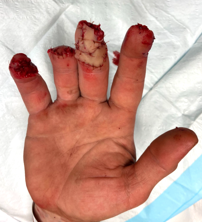
Figure 10: S/p repair.
Discharge Instructions:
- Keep dressing on until clinic follow-up
- Keep the injured hand elevated as much as possible to aid with resolution of swelling
- Keflex 1 week
- Follow-up with Hand Clinic in 1 week
Follow Up:
- IF and SF: Granulating wounds
- LF: Partial take of skin graft along the edges
- RF: healing appropriately
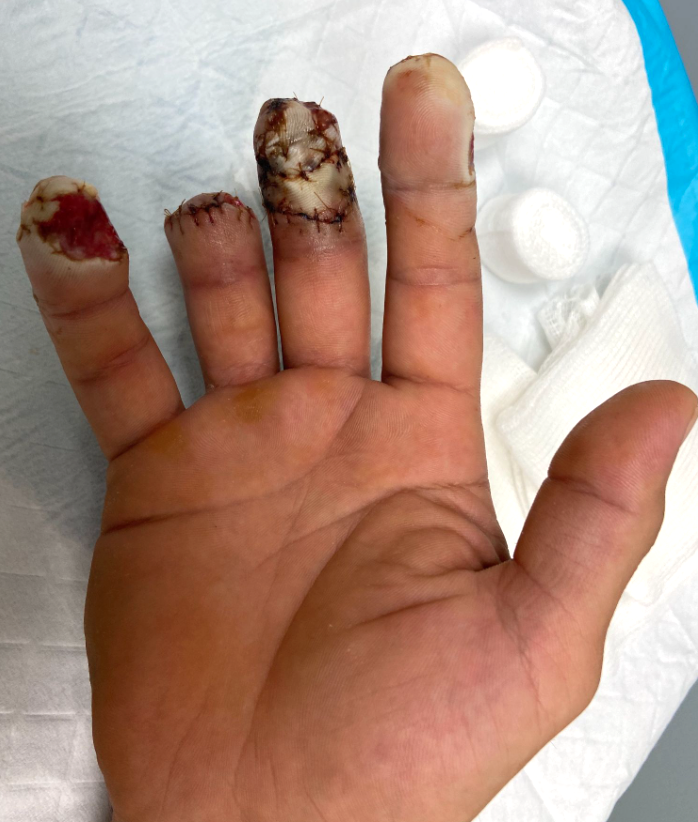
Figure 11: s/p 1 week.
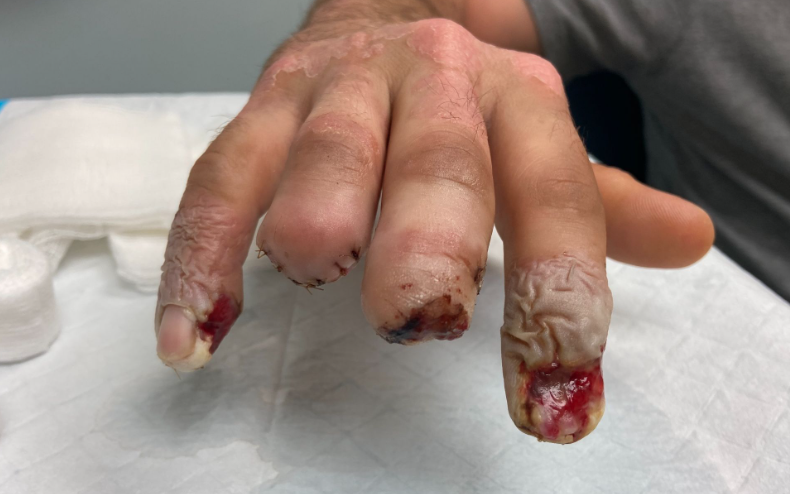
Figure 12: s/p 1 week.
2. 1-month post-injury: All digits healing appropriately. Dressing changed to daily Bacitracin and Band-Aid.
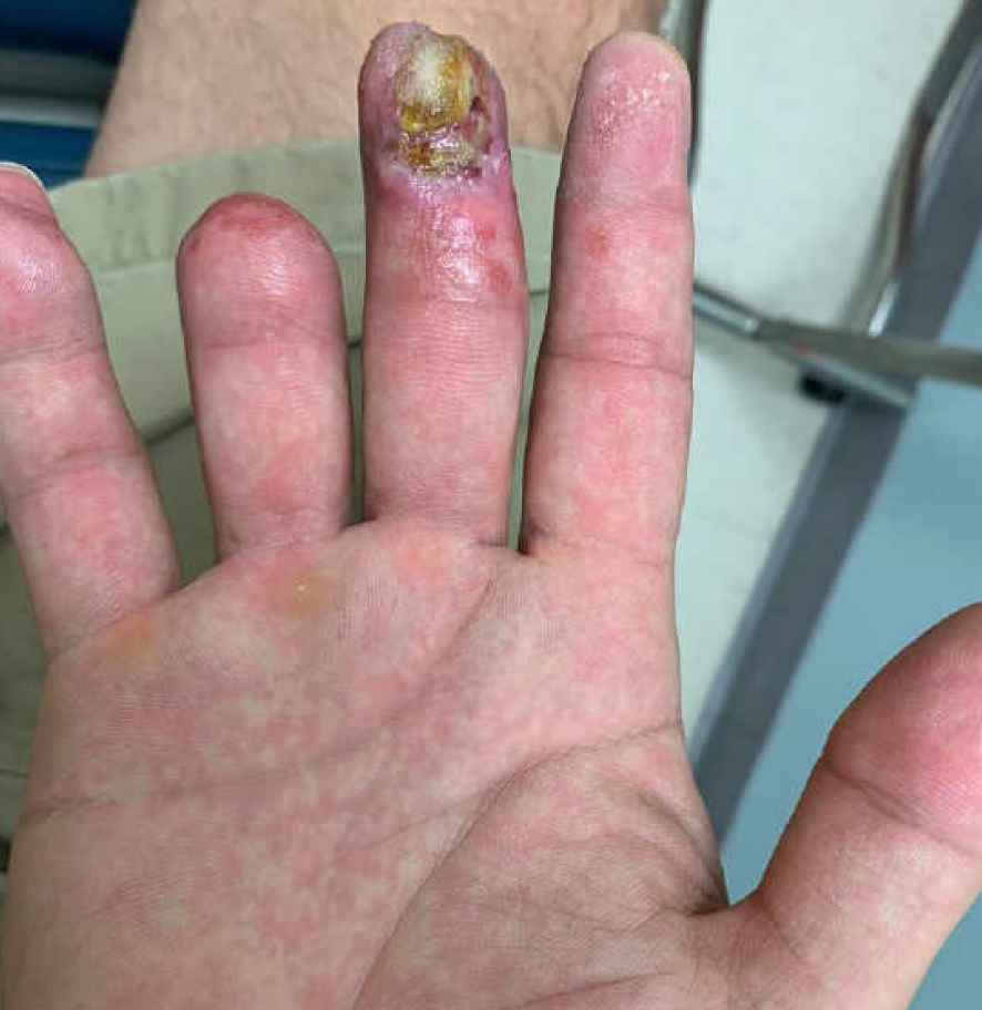
Figure 13: s/p 1 month.
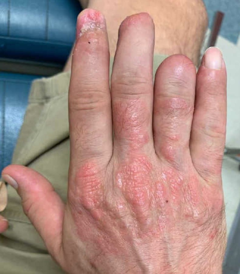
Figure 14: s/p 1 month.

Figure 15: s/p 1 month.
3. 3-month post-injury: Improved wound healing to all digits with resolving tingling to LF. Plan is to return to clinic in 3 months to discuss further treatment. Diligent hand therapy encouraged in the interim.
