Findings:
7cm linear scar, axially oriented along the left proximal lateral leg without any remaining palpable mass
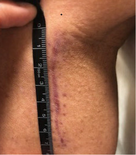
A. Preoperative: Left proximal lateral leg with healed excisional biopsy scar
Treatment:
No evidence of metastatic disease or remaining localized tumor on imaging. Patient underwent definitive excision of surrounding soft tissue and skin with placement of brachytherapy catheters and temporary NPWT closure. She completed a 5 day course of brachytherapy. Her final pathology revealed no residual tumor and all margins negative. She returned to the operating room with the plastic surgery team nine days later.
She was placed supine with a left lateral hip bump. Her final defect measured 13x6cm on the left proximal lateral leg, oriented vertically with muscle and subcutaneous tissue at the wound base. Due to her history of brachytherapy, it was determined that she would most benefit from fasciocutaneous flap reconstruction in order to reconstruct the defect with well perfused tissue. A hand-held doppler was utilized to identify perforators adjacent to the posterior margin of the defect. A Keystone fasciocutaneous flap was designed along the posterior aspect of the defect, incorporating the identified perforators, with the flap measuring 20x7cm.
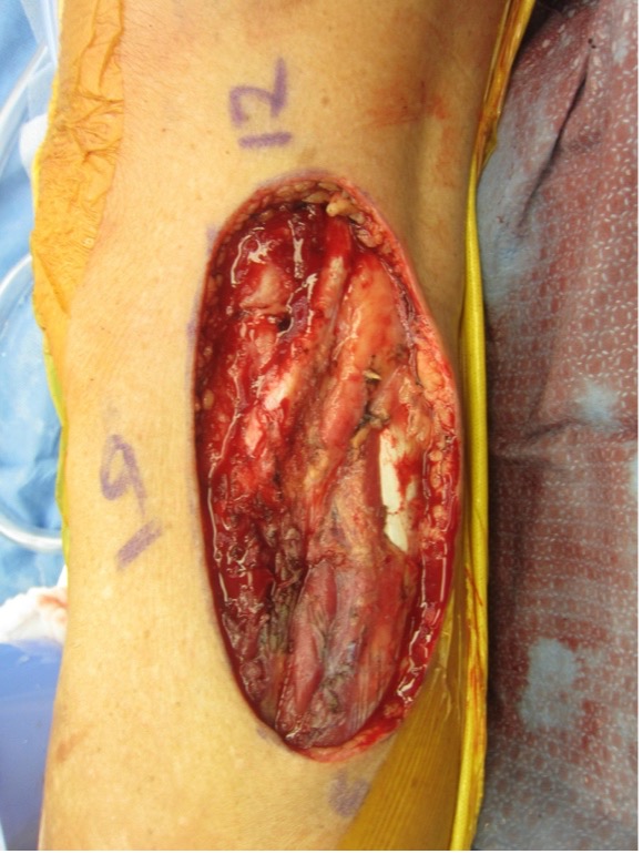
B. Intraoperative: left proximal lateral leg defect after definitive sarcoma excision and brachytherapy, 13x6cm
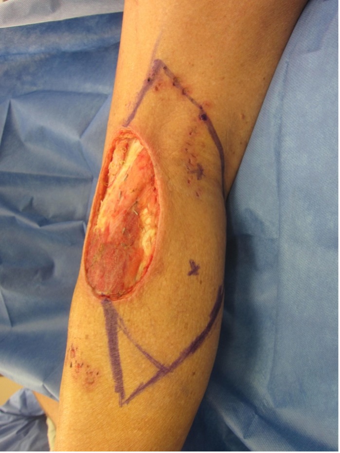
C. Intraoperative: Keystone fasciocutaneous flap design incorporating cutaneous perforators identified via hand held doppler (perforators marked by “x”)
The flap margins were incised through skin, subcutaneous tissue and deep fascia and then advanced into the defect and secured into place using interrupted deep dermal monocryl sutures and a running prolene on the skin. A 15Fr blake drain was placed under the flap and brought out distally. Attention was then turned to the donor site defect. The proximal and distal aspects of the defect were closed in a V-Y advancement fashion with interrupted deep dermal monocryl sutures. However, the central portion of the donor site defect was not able to close primarily without significant tension. Thus, the remaining wound edges were sutured down to the underlying muscle to create a uniform wound bed contour. A split thickness skin graft was obtained from the left lateral thigh, harvested at 0.012in using a dermatome, fenestrated and applied to the remaining donor site defect with chromic suture. A NPWT bolster was applied over the skin graft, along with a bulky dry dressing and left knee immobilizer.
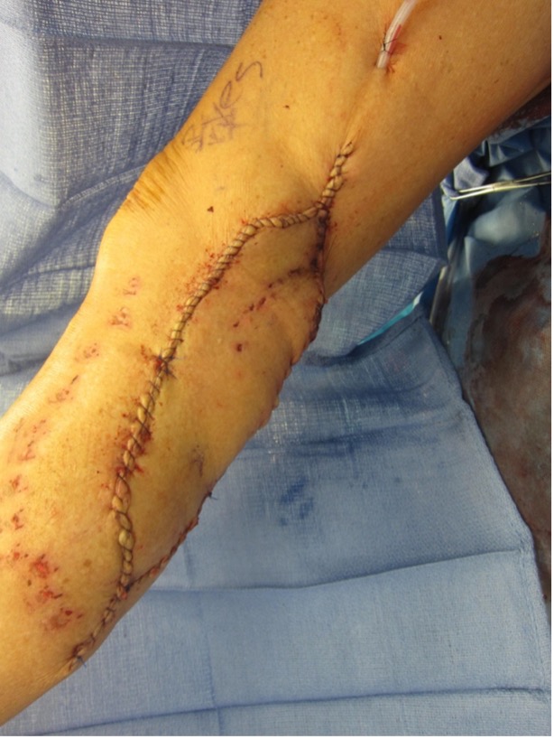
D. Intraoperative: Advancement of Keystone fasciocutaneous flap into defect of left proximal lateral leg defect and closure (note punctate scars of the skin, medial to the flap, consistent with prior brachytherapy catheter placement)
Follow Up:
Patient was allowed to ambulate with left knee immobilizer in place at all times postoperatively. The wound vac bolster was removed 1 week postoperatively with 80% graft take noted. Daily xeroform and dry gauze dressing changes were applied over the skin graft for 4 weeks until the areas of granulation tissue completely epithelialized through secondary intention. Sutures were removed 3 weeks postoperatively and the patient was allowed to ambulate without the knee immobilizer. Patient returned to all normal activity by 3 months postoperatively without any evidence of sarcoma recurrence.
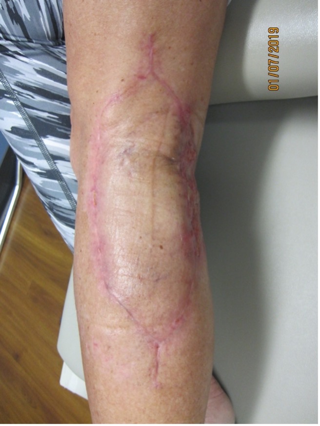
E. Postoperative: 2 month follow up with well healed incisions and epithelializing areas of split thickness skin graft loss at donor site
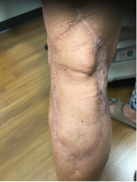
F. Postoperative: 3 month follow up with well healed incisions and donor site
