Lumbar Wound from Liposarcoma Resection

Brielle Weinstein
, Julian Pribaz

History:

The patient is a 59 year old female diagnosed with right lower back liposarcoma with positive margin after resection. She first noted the mass approximately one year prior to presentation to plastic surgery. The mass grew and initial excision was performed six months later. This had a positive deep muscular margin and she was then referred to a tertiary cancer care center. She completed radiation therapy prior to re-resection. In conjunction with the surgical oncology team, the patient presented to plastic surgery for resection of margin with reconstruction. She is otherwise healthy, non smoker, and active.

Findings:

After tumor extirpation, the plastic surgery team was presented with defect 17 x 9.5cm defect through skin, subcutaneous tissue, fascia over lumbar region. The patient had extensive radiation changes to the surrounding skin and soft tissue [Figure 1].

Lumbar Defect

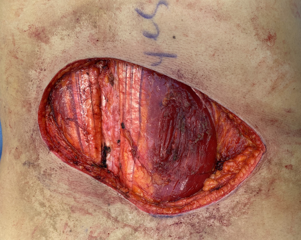
Figure 1. 17 x 9.5 cm defect through skin, subcutaneous tissue and fascia in lumbar region

Diagnosis:

Right lower back liposarcoma resection defect

Differential Diagnoses:

Pathology confirmed on presentation however prior to pathology differential diagnosis included lipoma, sarcoma

Workup Required:

 Patient did not require any specific imaging for plastic surgery portion of case

Plan:

Local fascio-cutaneous flaps for vascularized soft tissue coverage

Expertise Needed:

Plastic Surgeon

Treatment:

After thorough evaluation, the defect was reconstructed with a double, extended V to Y advancement. The underlying tissues were attached to the paraspinal ligaments for approximation and closure of dead space. The lateral and interior extensions allowed the flap to recruit tissue from three directions. As with any complex defect, the team began with creation of a template. We prefer to do this with Duoderm (ConvaTec, Bridgewater, NJ) as it best replicates the motion of soft tissues. We then designed the flap with a double V to Y design [Figure 2]. The flap was then incised through skin, subcutaneous tissue and fascia to adequately mobilize [Figure 3 and Figure 4]. Two surgical drains were placed and the wound was closed in a multi layer fashion. The deepest tissues were approximated to paraspinal fascia, subcutaneous tissue and deep dermis approximated with 2-0 vicryl sutures and the skin approximated with 3-0 nylon [Figure 5].
Lumber Defect with Flap Markings

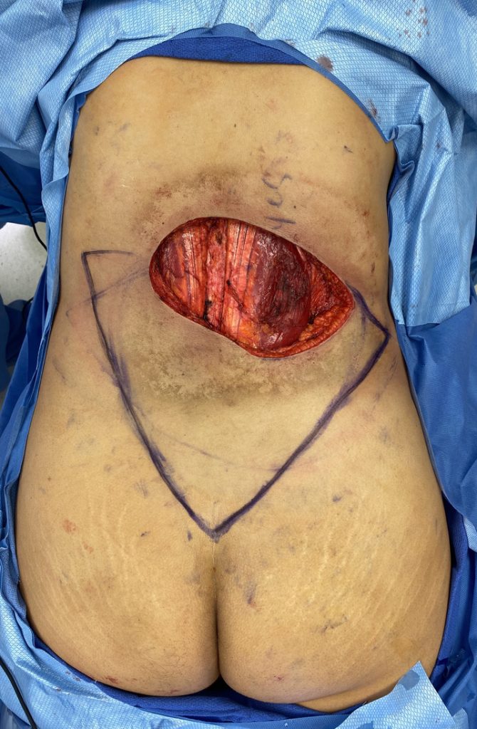
Figure 2. Flap design with double V to Y and three areas of advancement on bilateral flanks and gluteal cleft.

Lumbar Defect with Dissected Flap

Figure 3. Flap dissection through skin, subcutaneous tissue and fascia. Lateral mobility is noted on bilateral flanks and superior gluteal area.

Lumbar Defect with Mobilized Flap

Figure 4. Demonstration of mobility of lateral segments for medial soft tissue coverage.

Immediate Result

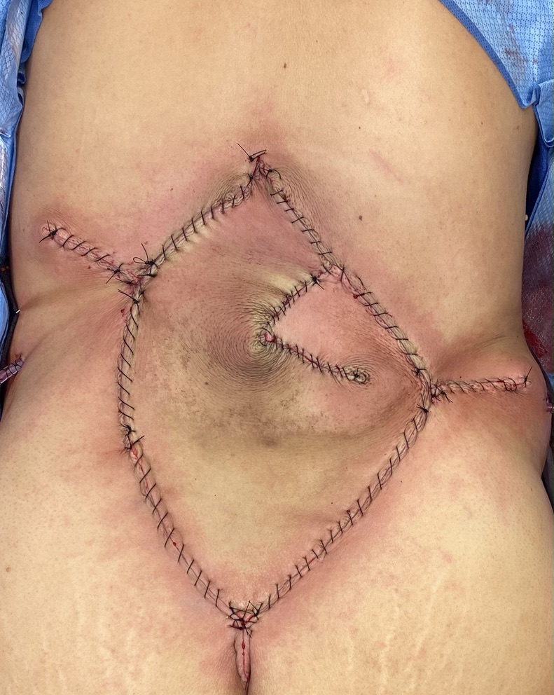
Figure 5. Immediate post operative result with double V to Y advancement with minimal tension closure in lumbar area.

Follow Up:

The patient has been seen in follow up at two weeks [Figure 6] for and two months [Figure 7] and is healing well. She has excellent truncal mobility.
Lumbar Reconstruction at Two Weeks

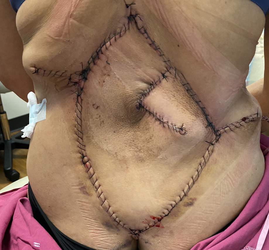
Figure 6. Post operative result at first follow up visit with well approximated incisions and no evidence of dehiscence, infection or drainage.

Lumbar Defect Reconstruction at Two Months

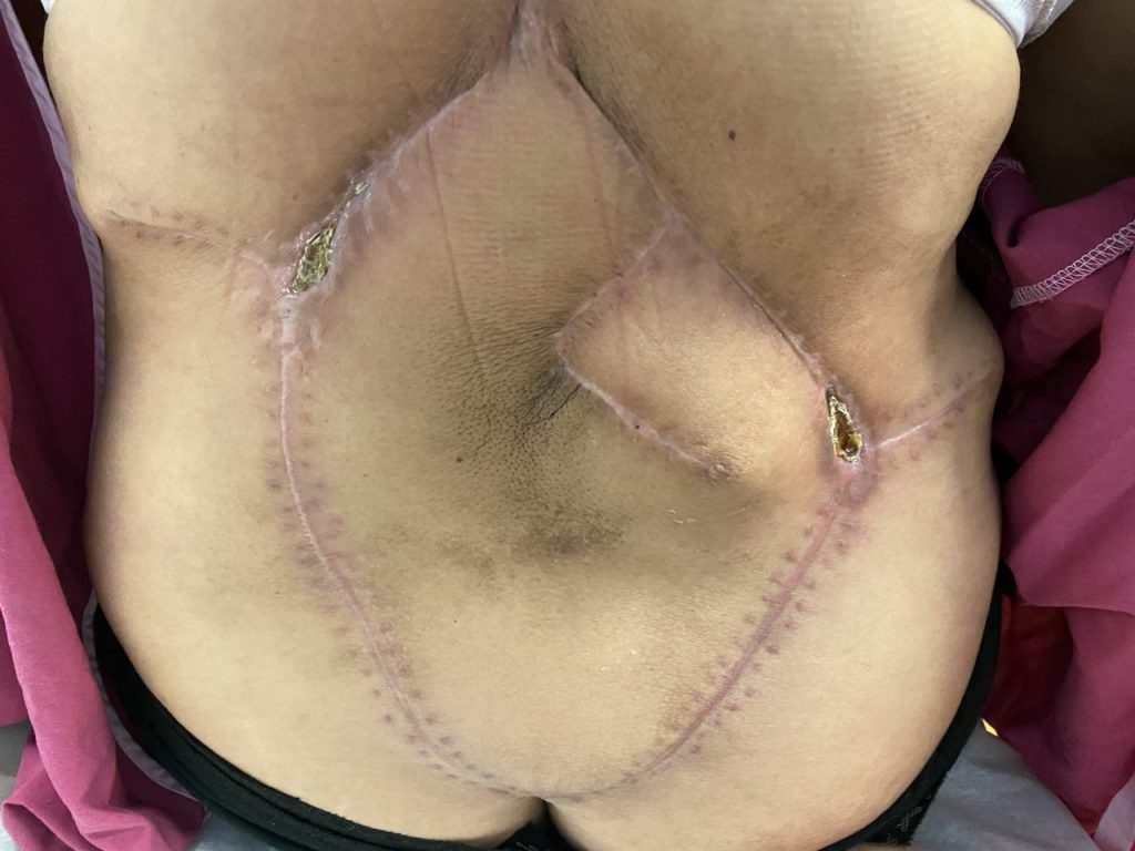
Figure 7. Two months follow up visit with well healed incisions and small areas of delayed wound healing at lateral T junctions

Print Friendly, PDF & Email

References

Pribaz JJ, Chester CH, Barrall DT. The extended V-Y flap. Plast Reconstr Surg. 1992 Aug;90(2):275-80. PMID: 1631219.
Pribaz JJ, Chan RK. Where do perforator flaps fit in our armamentarium? Clin Plast Surg. 2010 Oct;37(4):571-9, xi. doi: 10.1016/j.cps.2010.06.007. Epub 2010 Jul 29. PMID: 20816513.
Hernekamp JF, Cordts T, Kremer T, Kneser U. Perforator-Based Flaps for Defect Reconstruction of the Posterior Trunk. Ann Plast Surg. 2021 Jan;86(1):72-77. doi: 10.1097/SAP.0000000000002439. PMID: 32541540.
https://pubmed.ncbi.nlm.nih.gov/1631219/
https://pubmed.ncbi.nlm.nih.gov/20816513/
https://pubmed.ncbi.nlm.nih.gov/32541540/

Related Articles