Dog bite to lower eyelid

Mihail Climov, MD
, Kate Gelman
, Sebastian Brooke, MD

History:

A generally healthy 26-year-old male presented to the emergency room due to a right lower eyelid accidental laceration while playing with his dog. Before presenting to the emergency room, he went to an Urgent Care Clinic, where he received a tetanus booster and was started on Augmentin. There was no bleeding, and the wound appeared clean; the patient did not report any vision problems or sensory changes. The dog was a vaccinated German Shepard with no history of prior attacks.

Findings:

15 x 5 mm curved, clean, full-thickness laceration to the right lower lid, laterally to the tear trough ligament with malar fat compartment exposure. The wound appeared clean, with several blood clots and a few rough edges. No acute bleeding was present. No eye movement restriction, no sensation changes, nor vision changes. No evidence of fractures or signs of infection.

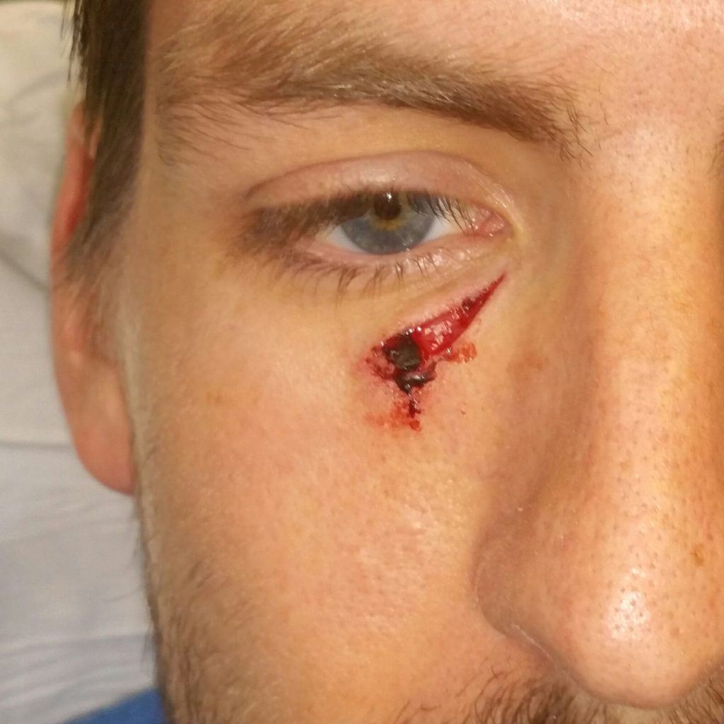
Figure 1. Right lower eyelid laceration due to a dog bite


Diagnosis:

Acute, traumatic full-thickness skin laceration to the right lower eyelid due to accidental dog bite in a healthy 26-year-old male without fracture or nerve injury.

Differential Diagnoses:

Fractures, injuries, or ocular injuries were ruled out.

Workup Required:

Clinically due to the superficiality of the wound, no concerning symptoms such as pain, vision changes, tenderness on palpation, bony step-offs, or Battle sign – the workup consisted of taking a thorough history of the details of the event, on the vaccination status of the dog and tetanus booster history. If we had any concerns, we would require a CT scan to assess the extent of injury, potential fractures, or retained teeth, and an ophthalmology consult would have been called in case of vision changes or epiphora.

Plan:

  1. Ensure that the status of the dog (own/unknown) and whether it was vaccinated
  2. Ensure patients’ tetanus vaccination is up to date or administer a booster
  3. Assess for any vision changes, paresthesia/sensory changes
  4. Anesthetize with a local anesthetic
  5. Copious irrigation to clean the wound
  6. Inspect the wound, trim the edges, and close primarily with 6-0 Prolene, interrupted suture, and no glue due to the risk of infection
  7. Discharge home with one week of Augmentin and topical Bacitracin to the incision
  8. Sutures removal in 5-7 days
  9. Follow up in 5-7 days and then when needed


Expertise Needed:

Emergency Room MD, PA, NP, or RN


Treatment:

The skin around the laceration was cleaned with Chloroprep carefully not to get into the eye or the wound (safer alternatively for the periorbital area would be Betadine). Anesthesia was performed with one ml of 1% lidocaine with epinephrine. The site was draped in a sterile fashion. Once anesthetized, copious irrigation of the wound with saline was performed. The wound was explored, and no foreign bodies were found; the rugged wound edges were trimmed with sharp iris scissors to obtain healthy, smooth wound margins. The wound was primarily closed with 6-0 Prolene sutures in an interrupted fashion2, spaciously allowing the wound to drain in case of exudate. The patient tolerated the procedure very well and was pleased with the result. He was discharged with instructions to carefully monitor for any signs of infection, to continue Augmentin for a total of one week, to apply Bacitracin to the wound daily, and follow up for suture removal in 5-7 days3,4.

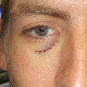
Figure 2. Right lower eyelid laceration after washout and repair


Follow Up:

By two weeks, the incision was completely healed.

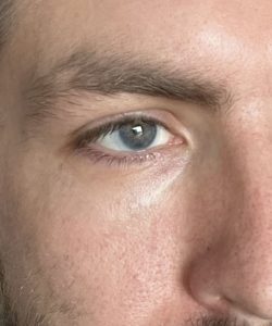
Figure 3. Three years after the repair

Print Friendly, PDF & Email

References

Sadiq MAA, Corkin F, Mantagos IS. Eyelid lacerations due to dog bite in children. J Pediatr Ophthalmol Strabismus. 2015;52(6):360-363. doi:10.3928/01913913-20150901-02
Bullocks J, Hsu P, Izaddoost S, Hollier L. Plastic Surgery Emergencies. Principles and Techniques. Second edi. New-York: Thieme; 2017.
Wu PS, Beres A, Tashjian DB, Moriarty KP. Primary repair of facial dog bite injuries in children. Pediatr Emerg Care. 2011;27(9):801-803. doi:10.1097/PEC.0b013e31822c1112
https://pubmed.ncbi.nlm.nih.gov/26371465/
https://www.thieme-connect.de/products/ebooks/lookinside/10.1055/b-0037-144881
https://pubmed.ncbi.nlm.nih.gov/21878832/

Related Articles