Findings:
77 year old woman with a 6×2 cm ulcer in the left axilla. The ulcer showed what appeared to be hyper granulation.
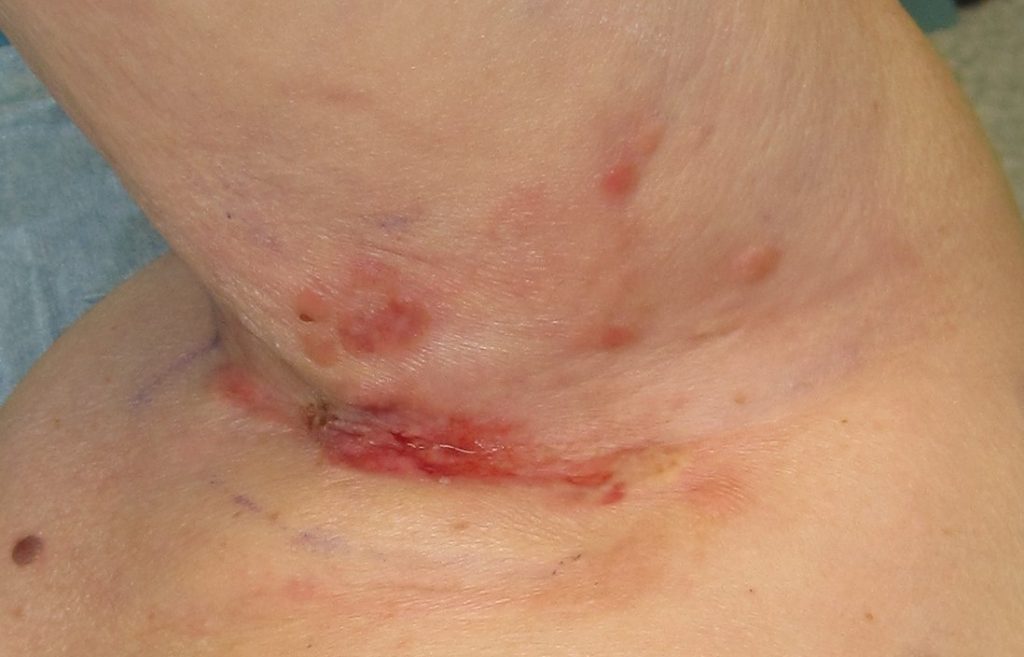
Fig.1. Left armpit showing central ulcer surrounded by nodular skin lesions, highly suspicious of representing recurrent breast cancer.
Treatment:
Wide local excision and closure with a fascio-cutaneous transposition flap incorporating thoracodorsal perforators marked with a hand held doppler.
A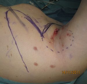
B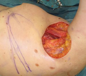
C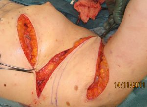
D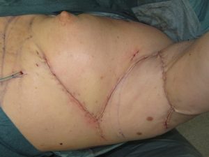
Fig.2. A Preoperative markings. 3 thoracodorsal perforators identified with doppler and marked with dots.
B Wound defect after wide local excision. Pectoralis major muscle (upper part of wound) and latissimus muscle (lower part of wound) are visible.
C Flap raised and transposed into defect.
D End of procedure. Flap healed at 2 week follow up without necrosis or infection.
