Lip Laceration Extending Through Vermillion

Seth Fruge M.D.
, Bohdan Pomahac M.D.

History:

A 74-year-old female with no past medical history presented to the emergency department after a fall from standing while in her home. She reports that she tripped over a vacuum cord and struck her face on the ground. She denies any loss of consciousness or any other injuries. She reports minimal bleeding from her central lip that was controlled with pressure. Patient was up to date on her T-DAP. No further imaging or labs were obtained by the emergency department. The Plastics and Reconstructive Surgery team was consulted for the lip laceration as it crossed the vermillion border.

Findings:

On exam, there was a 2.5cm vertically oriented laceration located just medial to the right philtral column that crossed 1 cm though the vermillion border and into the wet lip mucosa. There was a central stellate portion of this laceration at the vermillion border in the central portion of cupids bow. Orbicularis Oris muscle was visible along the superior portion of the laceration, but the muscle was not violated. There was no loss of teeth or evidence of facial fractures.

Diagnosis:

Complex laceration of upper lip including vermillion border.

This image illustrates the wound on presentation. Complex laceration of upper lip including vermillion border

Fig. 1. This image illustrates the wound on presentation. Complex laceration of upper lip including vermillion border

Differential Diagnoses:

Not applicable.

Workup Required:

Cranial Nerves 2-11 were tested and were without deficit. No intra-oral lacerations, malocclusion, or loose teeth were noted on exam. Verbal consent was then obtained for irrigation, debridement, and closure of her lip laceration.

Plan:

Repair of the laceration under local anesthesia in the Emergency Department.

Expertise Needed:

Plastic Surgeon

Treatment:

The patient was placed in the supine position with good lighting. The vermillion border on both sides of the wound was marked with a surgical marker prior to infiltration with local anesthetic. The wound was infiltrated locally with 5cc of 1% xylocaine with epinephrine (1:100000). Direct infiltration was chosen over an infra-orbital nerve block as the wound was central and was not very large. The wound was irrigated thoroughly with 1L of sterile saline and examined for depth and involvement of surrounding structures. As aforementioned, the orbicularis oris muscle was intact and did not require repair. The oral mucosa was also not violated which was also consistent with the prior exam. The vermillion border was first using the prior surgical markings and a 7.0 prolene simple interrupted suture. The skin along the right philtral column and along the dry lip was also closed using simple interrupted 7.0 prolene sutures. At the transition point of the wet lip and dry lip, a simple interrupted 5.0 chromic gut suture was used. Simple interrupted 5.0 chromic gut sutures were also used to close the remainder of the wet lip laceration. Bacitracin was applied to the wound and the patient was instructed to apply Bacitracin ointment twice daily until follow up. She was also instructed to avoid sun exposure and use SPF-30 or higher lotion if exposed to sun for the next year. This was done in order to decrease the chance of differential pigmentation of her scar. She was scheduled for follow up in 1 week for suture removal. On follow up visit, her wound was healing well, the sutures were removed, and the patient was instructed to apply Vaseline to the healed laceration until the lip was no longer crusting.

This image illustrates the wound after placing the sutures. The patient was scheduled for follow up in 1 week for suture removal.

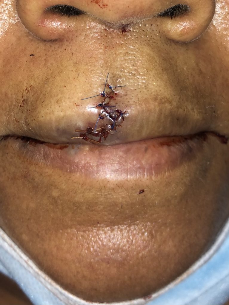
Fig. 2. This image illustrates the wound after placing the sutures. The patient was scheduled for follow up in 1 week for suture removal.

Follow Up:

One week for removal of non-resorbable sutures, followed by 1 month follow up for healing. Additional follow ups as needed depending on scar maturation and healing.
The healing wound after removal of the sutures and imaged during a follow up visit.

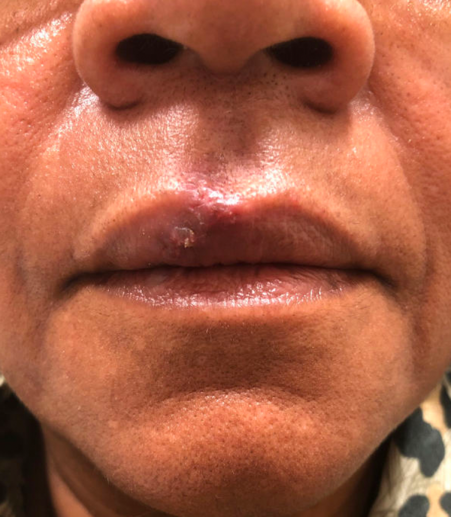
Fig. 3. The healing wound after removal of the sutures and imaged during a follow up visit.

Print Friendly, PDF & Email

References

Lip and Perioral Trauma. Grunebaum LD, Smith JE, Hoosien GE. Facial Plast Surg 2010 Dec 17; 26(6):433-44
Advanced Laceration Management. Brown DJ, Jaffe JE, Henson JK. Emerg Med Clin North Am 2007 Feb 25(1):83-99
Cost Effective Task Trainer for Complex Lip Laceration Repair. Walsh R, Lei C, Heimiller J, Sikon J, Palm KH. Cureus 2021 Mar 2;13(3):e13659. Epub 2021 Mar 2
http://dx.doi.org/10.1055/s-0030-1267717
http://dx.doi.org/10.1016/j.emc.2006.11.001
http://dx.doi.org/10.7759/cureus.13659

Related Articles