Infected ischemic diabetic foot ulcer

Miki Fujii MD
, Rica Tanaka MD

History:

A 82-year-old woman with history of type 2 diabetes mellitus and percutaneous coronary intervention was referred to our university hospital with left infected foot ulcer. She kept walking because she didn’t feel any pain. She developed a small ulcer on lateral side of the left fourth toe when she stumbled one month ago. The wound became larger, her foot was swollen, and the IV toe became necrotic.
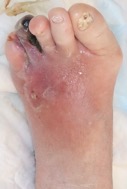
Findings:

There were wounds of the left fourth and fifth toe. The fourth toe was partially necrotic. The left foot was red, hot and swollen. Left dorsal pedis and posterior tibial artery were not palpable.

Diagnosis:

Infected ischemic diabetic foot ulcer

Differential Diagnoses:

Venous leg ulcer, pressure ulcer, vasculitis, skin cancer, pyoderma gangrenosum

Workup Required:

  1. ABI, SPP for arterial flow
  2. Assessment of arterial flow with ultrasound, contrast CT, or MRA
  3. Culture from the deep area of the ulcer
  4. Laboratory test including inflammatory markers and HbA1c

Plan:

  1. Hospitalization
  2. Infection control by antibiotics and bed rest
  3. Revascularization
  4. Debridement
  5. Wound bed preparation
  6. Reconstruction

Expertise Needed:

Wound specialist, Wound nurse, Endovascular Surgeon, Vascular surgeon, Radiologist, Diabetologist, Ultrasound technician

Treatment:

After emergency hospitalization, culture from the deep area of the ulcer was taken and antibiotics treatment was started. Antibiotics was performed empirically fist and changed to the most sensitive one based on the culture results. ABI was 0.8 of right and 0.44 of left. Contrast CT and angiography revealed the arterial occlusion of common femoral artery (CFA).

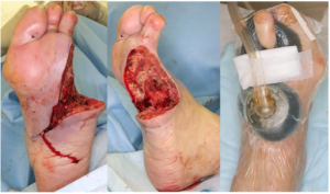
After revascularization by endarterectomy of CFA, emergency debridement was performed and left the wound open. 7 days later, additional debridement was performed and applied Negative pressure wound therapy with instillation and dwell time;NPWTi-d ( 3M™ V.A.C. ®️ Ulta Therapy System; 3M). After 3 weeks, infection was completely controlled and wound bed preparation was completed. Mesh skin graft was performed and wound was healed without any complication.
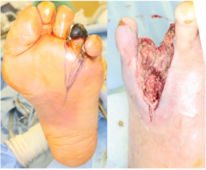
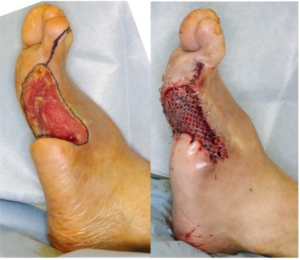
Follow Up:

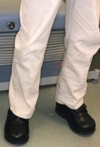
After 2 years the wound had stayed healed and she could walk with custom made foot ware. There was no recurrence.

Print Friendly, PDF & Email

References

Conte MS, Bradbury AW, et al.; GVG Writing Group.Global vascular guidelines on the management of chronic limb-threatening ischemia. J Vasc Surg. 69(6S):3S-125S.e40, 2019
Senneville É, Albalwi Z, et al.; International Working Group on the Diabetic Foot (IWGDF). Guidelines on the diagnosis and treatment of foot infection in persons with diabetes (IWGDF 2023 update)
Mills JL Sr, Conte MS, Armstrong DG, et al; Society for Vascular Surgery Lower Extremity Guidelines Committee. The Society for Vascular Surgery Lower Extremity Threatened Limb Classification System: risk stratification based on wound, ischemia, and foot infection (WIfI). J Vasc Surg. 59(1):220-34.e1-2, 2014
https://pubmed.ncbi.nlm.nih.gov/31159978/
https://iwgdfguidelines.org/guidelines-2023/all-guidelines-2023/
https://pubmed.ncbi.nlm.nih.gov/24126108/

Related Articles