Forehead/Glabella laceration

Olivier F. Noel, M.D., Ph.D.
, Melissa Mastroianni, M.D.

History:

A 67 year old female with deep forehead and glabella laceration sustained after fall onto a tree stump. She denied any loss of consciousness or any other injuries. She reported significant bleeding from her forehead that was controlled with pressure in the ED. Patient was up to date on her T-DAP. No further imaging or labs were obtained by the emergency department. The Plastic and Reconstructive Surgery team was consulted for the forehead/glabella laceration.

Findings:

On exam, deep  laceration to forehead extending down into glabella region and avulsed, elevated skin flap without tissue loss. Moderate continuous oozing from deeper soft tissue. Significant left periorbital swelling and ecchymosis. There were no other injuries or evidence of facial fractures.

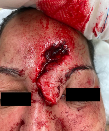
Figure 1. Shows deep laceration to forehead extending to glabella region.


Diagnosis:

Complex laceration of forehead and glabella regions. No nasal, orbital or other facial fractures.

Differential Diagnoses:

 Fractures and concussion which have been ruled out.

Workup Required:

Cranial Nerves 2-11 were tested and were without deficit. No nasal, orbital, or forehead fractures, or intraoral lacerations, malocclusion, or loose teeth were noted on exam. Verbal consent was then obtained for irrigation, debridement, and closure of his lip laceration.

Plan:

Repair of the laceration under local anesthesia in the Emergency Department.

Expertise Needed:

Plastic Surgeon.

Treatment:

The patient was placed in the supine position with good lighting. The wound was copiously irrigated with saline and betadine solution. In the muscle, 4-0 Monocryl sutures were used. In the superficial layer, 6-0 Monocryl dermal sutures were used to approximate the dermal edges. 6-0 Prolene interrupted sutures were used for the best skin approximation. Bacitracin was applied to the wound and the patient was instructed to apply Bacitracin ointment twice daily until follow up. She was also instructed to avoid sun exposure and use SPF-30 or higher lotion if exposed to sun for the next year. This was done in order to decrease the risk of hyperpigmentation of her scar. She was scheduled for follow up in 1 week for suture removal. She received 5 days of prophylactic antibiotics. On follow up visit, her wound was healing well, the sutures were removed, and the patient was instructed to apply Vaseline to the healed laceration until the wound was no longer crusting.

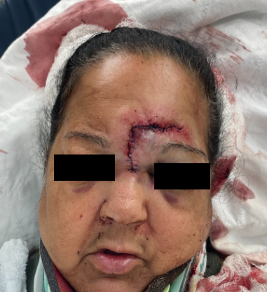
Figure 2. Shows repaired laceration.

Follow Up:

One week for removal of non-resorbable sutures, followed by 3 weeks follow up for healing. Additional follow ups as needed depending on scar maturation and healing. She was told that she would likely need a revision of her depressed scar.

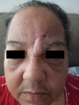
Figure 3. One month follow up.

Print Friendly, PDF & Email

References

Bhattacharya V. Management of soft tissue wounds of the face. Indian J Plast Surg. 2012 Sep;45(3):436-43. doi: 10.4103/0970-0358.105936. PMID: 23450264; PMCID: PMC3580340.
Yaghoubian JM, Aminpour S, Anilus V. Supertrochlear Nerve Block. 2022 Jun 27. In: StatPearls [Internet]. Treasure Island (FL): StatPearls Publishing; 2022 Jan–. PMID: 32644465.
Napier A, De Jesus O, Taylor A. Supraorbital Nerve Block. 2022 May 29. In: StatPearls [Internet]. Treasure Island (FL): StatPearls Publishing; 2022 Jan–. PMID: 30725622.
https://www.ncbi.nlm.nih.gov/pmc/articles/PMC3580340/
https://pubmed.ncbi.nlm.nih.gov/32644465/
https://pubmed.ncbi.nlm.nih.gov/30725622/

Related Articles