
History:
A 55 year old male with no significant past medical history presented to the emergency department, after sustaining a severe lawnmower injury to his right foot. The patient had slipped under an operating mower.
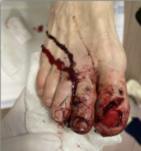
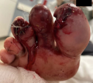
Emergency department
Findings:
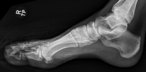
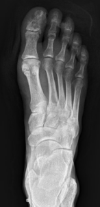
Initial imaging
– Acute, open, displaced fractures of the right 1st, 2nd, and 3rd toe phalanges.
– Lacerations of the right 1st, 2nd, and 3rd toes.
– Soft tissue necrosis and focal flap necrosis of the right foot status post debridement and treatment of laceration
The patient underwent debridement and repair of lacerations with podiatry in the operating room. He was discharged with an oral antibiotic course. Upon his follow up appointment, the patient reported intermittent shooting pain at the site. Clinical examination of the surgical site demonstrated intact sutures with no signs of acute infection or purulent drainage. However, necrotic changes were appreciated.
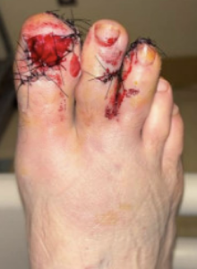
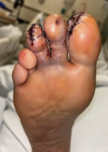
Post-op day 1
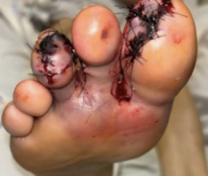
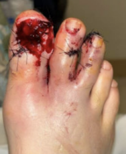
Post-op day 2
Diagnosis:
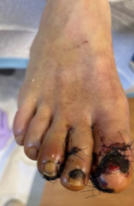
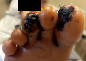
Post-op week 2
Treatment:


Session 5/40 of HBOT
At approximately post-operative month 1, the patient went for follow up appointment with the podiatrist. Repeat X-rays showed healing highly comminuted fracture of the 1st distal phalanx, 2nd proximal, middle and distal phalanges and 3rd distal phalanx in similar alignment. Weight-bearing status changed to weight bearing as tolerated with a flat surgical shoe with modifications. The remaining sutures were removed.
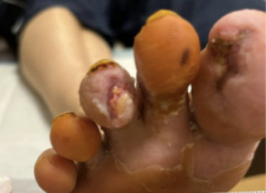
Session 13/40 of HBOT
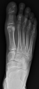
Post-op month 1
Follow Up:
He completed 40 hyperbaric oxygen therapy sessions and tolerated the sessions well.
Repeat X-ray shows multiple healing fractures of the 1st through 3rd toes. Patient was congratulated on achieving full healing with no dressing indicated. At post-op month 3, return to normal activities with no restrictions was recommended.
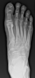
Post-op month 2

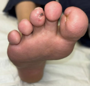
Post-op month 2
