Fingertip amputation with exposed bone in 4-year old

Olivier F. Noel, M.D., Ph.D.
, David Colen, M.D.

History:

A 4 year old male with no past medical or surgical history presented to the emergency department (ED) after his right middle finger got stuck on a door hinge and sustained an amputation of the fingertip with bone exposed. The fingertip was brought to the ED in a plastic bag being cooled on ice. Patient was up-to-date on his TDAP vaccinations. There were no child abuse/neglect concerns. Hand and dedicated finger X-Rays were obtained in the ED. The Plastic and Reconstructive Surgery team was consulted for management of the fingertip amputation.

Findings:

Right middle finger with distal tip amputation. The distal phalanx was exposed but appeared intact. Patient was able to flex and extend the digit at the DIP joint. Hemostasis was achieved in the ED with compression. There were no other injuries.

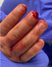
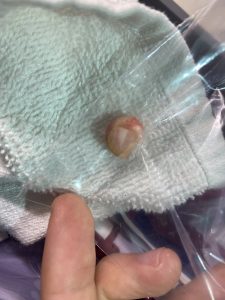
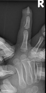
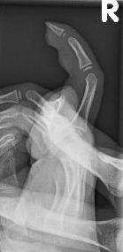
Figure 1A-E. Shows amputated right middle fingertip, the amputated tip and XRays upon arrival to the ED.

Diagnosis:

Right middle fingertip amputation with exposed bone but no fracture.

Differential Diagnoses:

Not applicable

Workup Required:

X-rays of the right hand to evaluate for fractures and possible retained foreign bodies.

Plan:

Repair of right fingertip amputation under conscious sedation in the Emergency Department.

Expertise Needed:

Plastic surgeon or Hand surgeon.

Treatment:

Local anesthesia with 1% plain lidocaine provided a digital block. The hand was prepped with betadine and draped using sterile towels. The wound as well as the amputated fingertip were copiously irrigated with normal saline and betadine. No functional structures were injured and the distal phalanx was intact. Picture 1 demonstrated the amputated digit upon arrival to the ED. The amputated fingertip was reapproximated to its anatomical position with simple interrupted 5-0 Chromic sutures as shown in Figure 2. A bulky dry soft dressing was then applied for 2 weeks to keep sutures in place, reduce involuntary movement and tension on the wound and prevent the pediatric patient from disrupting the healing process.

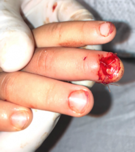
Figure 2. Shows the amputated finger reapproximated to anatomical position using 5-0 Chromic sutures.

Follow Up:

The patient was seen two weeks later in clinic. At the time, the digit was demonstrating healing with expected ischemic changes. Recommended bacitracin and dressings changes for protection and comfort.

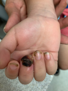
Figure 3. Two week follow up

When he was seen 6 weeks after the injury, the fingertip had  healed and new nail growth was seen. There was no pain to the distal fingertip and he was able to use the hand without limitations.

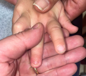
Figure 4. Six week follow up

Print Friendly, PDF & Email

References

Loewenstein SN, Adkinson JM. Pediatric Fingertip Injuries. Hand Clin. 2021 Feb;37(1):107-116. doi: 10.1016/j.hcl.2020.09.009. PMID: 33198910.
Rosenthal LJ, Reiner MA, Bleicher MA. Nonoperative management of distal fingertip amputations in children. Pediatrics. 1979 Jul;64(1):1-3. PMID: 450537.
Sullivan MA, Cogan CJ, Adkinson JM. Pediatric Hand Injuries. Plast Surg Nurs. 2016 Jul-Sep;36(3):114-20. doi: 10.1097/PSN.0000000000000145. PMID: 27606586.
https://pubmed.ncbi.nlm.nih.gov/33198910/
https://pubmed.ncbi.nlm.nih.gov/450537/
https://pubmed.ncbi.nlm.nih.gov/27606586/

Related Articles