Eyebrow laceration

Olivier F. Noel, M.D., Ph.D.
, Omar Allam, M.D.
, Jackie Oh, M.D.

History:

A 74 year old female  sustained a left eyebrow laceration after a fall and hitting her head against a nightstand. She denied any loss of consciousness or any other injuries. She reported minimal bleeding from her eyebrow that was controlled with pressure. Patient was up to date on her T-DAP immunizations. No further imaging or labs were obtained by the emergency department. The Plastic and Reconstructive Surgery team was consulted for the eyebrow laceration.

Findings:

On exam, 3 cm laceration to left eyebrow extending down to the orbital rim. The eyelid had erythema and edema but was moving normally. There was no double vision and ocular movements were normal in all directions. Palpation of the entire supra- and infraorbital rims revealed no step off or other evidence of a fracture. There were no other injuries.

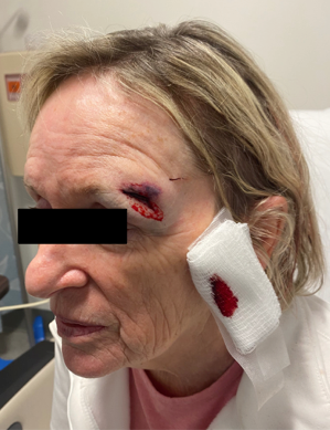
Figure 1. Shows laceration to the left eyebrow region.

Diagnosis:

Complex laceration of left eyebrow region. No evidence of fracture or intracranial injuries.

Differential Diagnoses:

None. Fracture and concussion have been ruled out.

Workup Required:

Cranial Nerves 2-11 were tested and were without deficit. No step off or area of focal tenderness palpated on exam. Consent was then obtained for irrigation, debridement, and closure of her eyebrow laceration.

Plan:

Repair of the laceration under local anesthesia in the Emergency Department.

Expertise Needed:

Emergency Room. MD, PA, NP or RN.

Treatment:

Wound gently cleaned with saline and betadine by gentle dabbing with dry sterile gauze. In the muscle, 4-0 Monocryl sutures were used. In the superficial layer, 6-0 Monocryl  sutures were used to approximate the dermal edges. 6-0 Prolene interrupted sutures were used for the best skin approximation. Bacitracin was applied to the wound and the patient was instructed to apply Bacitracin ointment twice daily until follow up. She was also instructed to avoid sun exposure and use sunscreen SPF-30 or higher lotion if exposed to sun for the next year. This was done in order to avoid hyperpigmentation of her scar. She was scheduled for follow up in 1 week for suture removal. She received 3 days of prophylactic antibiotics.

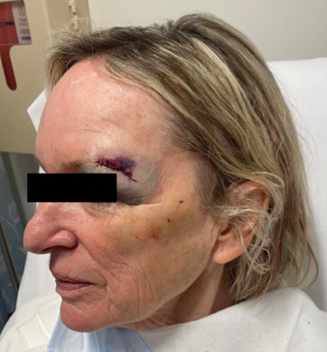
Figure 2. Shows repaired left eyebrow laceration.

Follow Up:

One week for removal of non-resorbable sutures, followed by 3 week follow up. Additional follow ups as needed depending on scar maturation and healing.

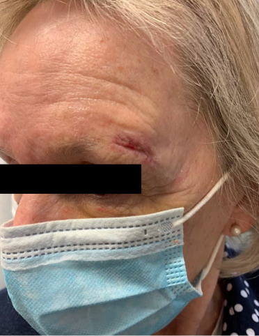
Figure 3. Three week follow up.

Print Friendly, PDF & Email

References

Brown DJ, Jaffe JE, Henson JK. Advanced laceration management. Emerg Med Clin North Am. 2007 Feb;25(1):83-99. doi: 10.1016/j.emc.2006.11.001. PMID: 17400074.
Bhattacharya V. Management of soft tissue wounds of the face. Indian J Plast Surg. 2012 Sep;45(3):436-43. doi: 10.4103/0970-0358.105936. PMID: 23450264; PMCID: PMC3580340.
Franz MG, Robson MC, Steed DL, Barbul A, Brem H, Cooper DM, Leaper D, Milner SM, Payne WG, Wachtel TL, Wiersema-Bryant L; Wound Healing Society. Guidelines to aid healing of acute wounds by decreasing impediments of healing. Wound Repair Regen. 2008 Nov-Dec;16(6):723-48. doi: 10.1111/j.1524-475X.2008.00427.x. PMID: 19128244.
https://pubmed.ncbi.nlm.nih.gov/17400074/
https://www.ncbi.nlm.nih.gov/pmc/articles/PMC3580340/
https://pubmed.ncbi.nlm.nih.gov/19128244/

Related Articles