Dorsal Hand and Wrist Wounds with Exposed Tendons

Bruce A. Kraemer, M.D.

History:

56-year-old female struck a deer while riding a motorcycle sustained in addition to a closed head injury, rib, scapular, left femur and transverse spinous process fractures along with an open dorsal wrist and hand wound.  Her other life threatening injuries took precedence over her hand wounds.

Findings:

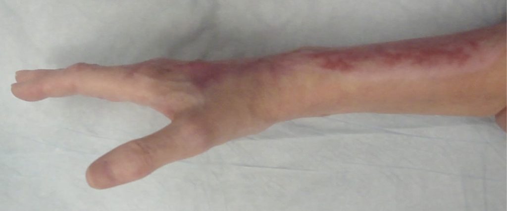
There was a 6 x 4 cm. tissue loss over the extensor carpi radialis longus and brevis tendons and the common digital extensor tendons at the wrist level as well a 2 x 2.5 cm wound involving the extensor tendons to the index finger over the index MCP joint.  Medically she was intubated and being treated for her severe closed head injury.

Fig.1. Dorsal Hand & Wrist Wound

Diagnosis:

Full thickness dorsal hand wound involving tendons in a patient with life threatening other injuries.

Differential Diagnoses:

None- Acute injury.

Workup Required:

X-rays to evaluate possible fractures and retained foreign bodies

Plan:

Initial wound management consisted of copious amounts of hydrogel covered with a polyurethane sheet dressing until the patient was stable to be taken to the operating room at post-injury day 5 for treatment of some of her other injuries.  At that point she had had multiple intravenous lines placed in her forearms, she had required pressor agents and she had developed a deep venous thrombosis of her left leg.  Given this the use of local transposition flaps were excluded and a rapid, simple but effective treatment option was desired.  The use of UBM-ECM wound devices was selected.

Expertise Needed:

A hand surgeon with particular interest in soft tissue reconstruction should assess the patient.  Knowledge of the use of Extracellular Matrix Wound Healing Devices is desired as these devices facilitate healing through a process termed “site-specific constructive remodeling”- promotion of the formation of more normal tissue which often minimizes scarring.  This M-2 macrophage directed healing is highly desirous in treating a seriously injured hand.

Treatment:

Operative placement of the UBM-ECM device was as follows: 1- 10 x 15 cm vacuum-pressed 6-layer sheets used as the outermost ECM layer, 2- 500 mg UBM-ECM powder is applied to the wound bed to initiate a robust constructive remodeling wound healing response, 3- 10 x 15 cm UBM-ECM lyophilized 2-layer sheet is then placed to “fill in” the remaining areas of tissue loss-  all of which is sewn in place (the 6-layer vacuum layer sheet is able to hold sutures) and then retained in place with petroleum impregnated gauze dressing.  A secondary dressing of IV tubing, plus a piece of hydroconductive wound dressing is placed under the outermost polyurethane sheet dressing.
Daily wound care consisted of addition of 5-10 cc saline via IV tubing under a secondary polyurethane sheet dressing.  She had her hand placed in a resting Orthoplast splint and had her arm elevated on 2 pillows.

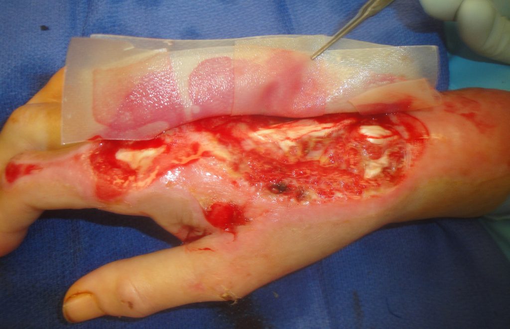
Fig.2. UBM-ECM Wound Device Placement 1

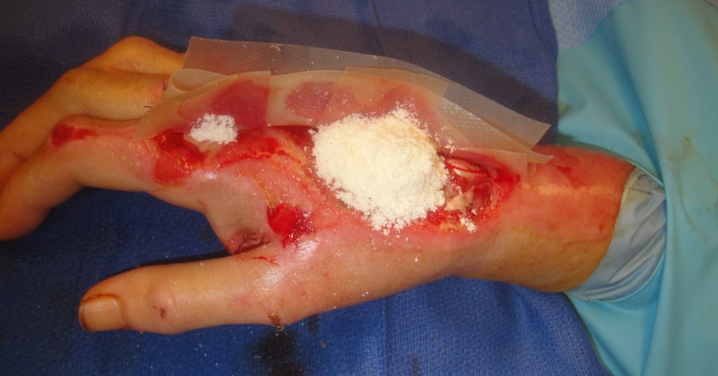
Fig.3. UBM-ECM Wound Device Placement 2

Fig.4. UBM-ECM Wound Device Placement 3

Fig.5. UBM-ECM Wound Device Placement 4

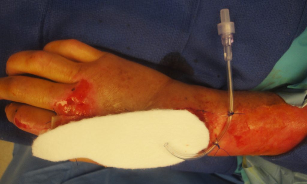
Fig.6. UBM-ECM Wound Device Placement Secondary Dressing

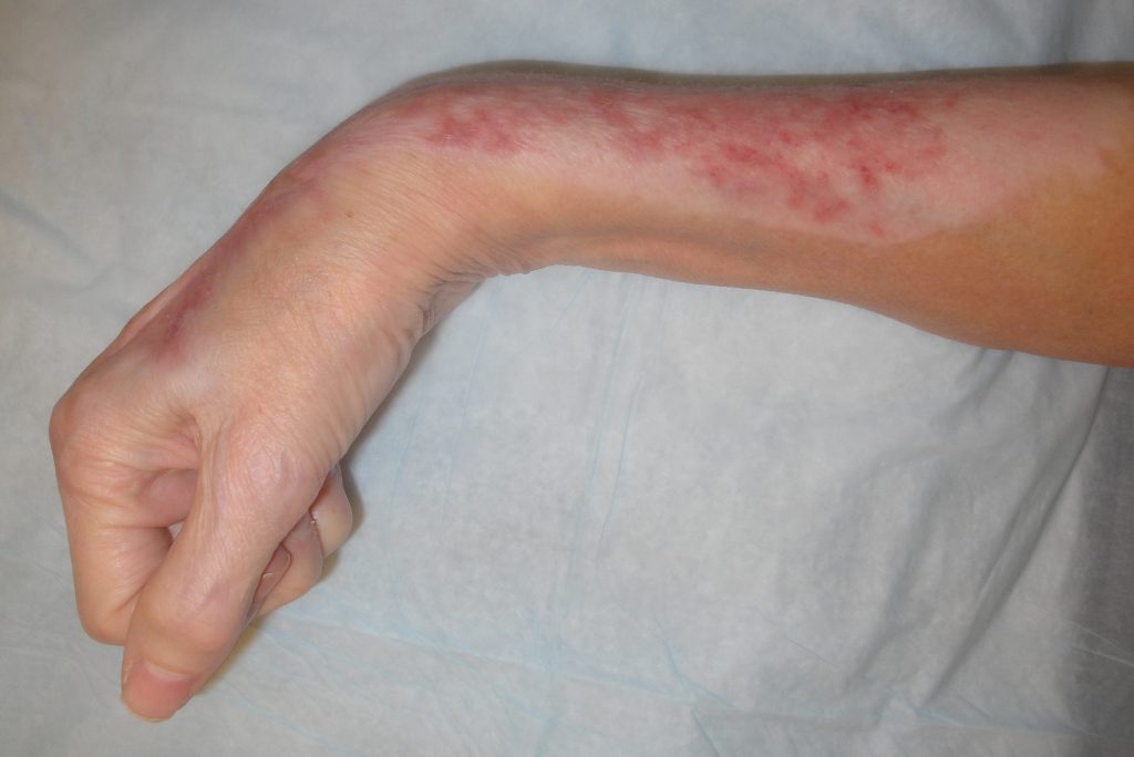
Fig.7. Postoperative wound appearance at week 3

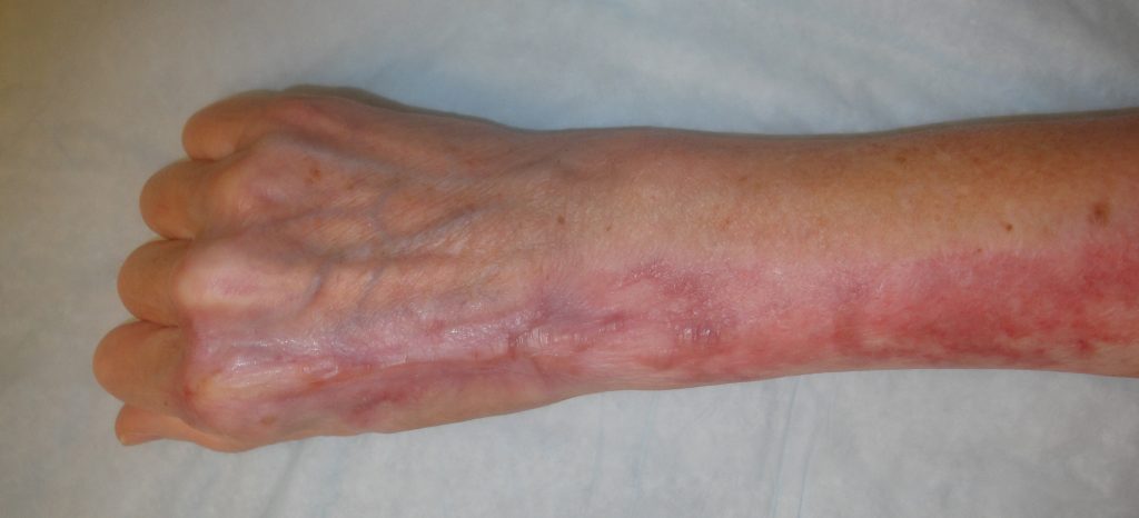
Fig.8. Final wound appearance 8 months post-op.

Final wound appearance 8 months post-op.

Final wound appearance 8 months post-op.

Final wound appearance 8 months post-op.

Follow Up:

She had minimal hand movement for the first 2 post-operative weeks after which she was continued in the splint until there was tissue coverage of her tendons at 3 months.  By 6 months she was able to follow simple verbal commands and was regaining normal use of her hand by nine months.  She showed no physical limitation of her hand use.
Print Friendly, PDF & Email

References

Geiger SE, Deigni OA, Watson JT, Kraemer BA. Management of Open Distal Lower Extremity Wounds With Exposed Tendons Using Porcine Urinary Bladder Matrix. Wounds- A Compendium of Clinical Research & Practice 2016;28(9):308-316.
Kraemer BA, Geiger SE, Deigni OA, Watson JT. Management of Open Distal Lower Extremity Wounds with Concomitant Fracture Using Porcine Urinary Bladder Matrix. Wounds- A Compendium of Clinical Research & Practice 2016; 28(11):387-394.
Behrens J, Kraemer BA. Abstract: Urinary Bladder Matrix-Extracellular Matrix (UBM-ECM) for Management of Complex Upper Extremity Wounds. Plast Reconstr Surg Global Open 2018;6(9 Suppl):51.
https://pubmed.ncbi.nlm.nih.gov/27701126/
https://pubmed.ncbi.nlm.nih.gov/27861131/
https://www.ncbi.nlm.nih.gov/pmc/articles/PMC6212145/

Related Articles