Findings:
Forehead: Abrasions over right forehead
Orbits: Small superficial laceration over right upper lateral eyelid. No periorbital ecchymosis, telecanthus, orbital tenderness, step-offs, or vertical dystopia.
Ophthalmology: No subconjunctival hemorrhage, no globe laceration/abrasion/rupture, no enophthalmos or exophthalmos; PERRLA; EOMI with no evidence of entrapment; vision and visual fields grossly intact
Nose: 4 cm diagonal laceration involving the upper and lower lateral cartilage (i.e. scroll area). Nasal bones were stable. No septal hematoma, no septal deviation.
Maxilla/Mandible: unremarkable
Sensation in involved areas was intact.
Facial motor exam: Normal and symmetric
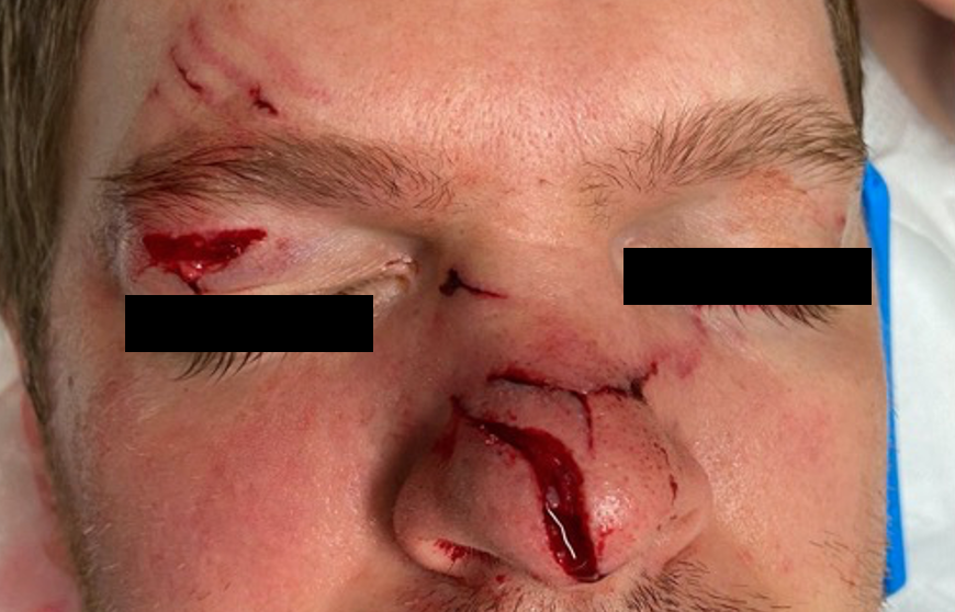
Figure 1: Zoomed out clinical image of all injuries- abrasion over right forehead, small superficial laceration over right upper lateral eyelid, and 4 cm diagonal laceration involving the upper and lower lateral nasal cartilage (i.e. scroll area).
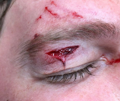
Figure 2: Abrasions over right forehead, along with small superficial laceration over right upper lateral eyelid.
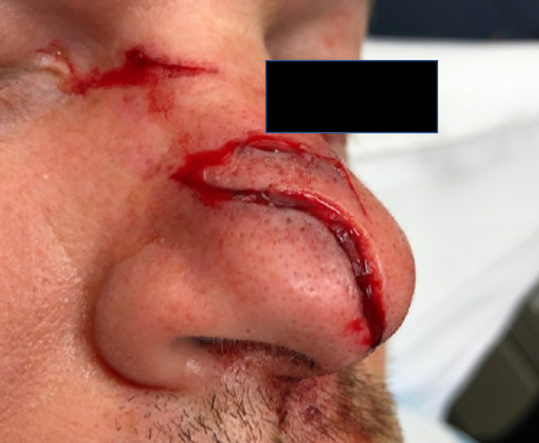
Figure 3: 4 cm diagonal laceration involving the upper and lower lateral nasal cartilage (i.e. scroll area).
Treatment:
Patient was placed in a supine position. Combination of local field block over right upper eyelid and nasal dorsum, and right infra-orbital nerve block (palpated intra-orbital foramen at mid-pupillary line and injected intra-orally) given with 5cc total of 2% lidocaine. Wound was thoroughly irrigated with normal saline. Then proceeded with approximation of upper and lower lateral cartilage to re-define scroll area with 5-0 Monocryl (buried simple interrupted). Remainder of skin (both nose and right upper eyelid) closed with 6.0 Prolene in simple interrupted fashion. Bacitracin was applied to wound. Patient tolerated the procedure well.
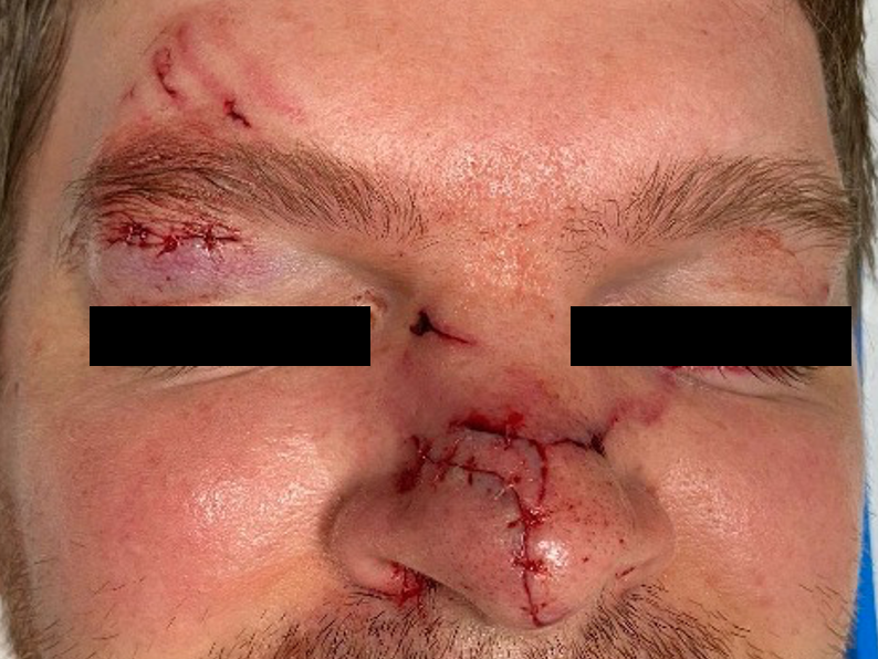
Figure 4: S/p bedside repair of linear right eyelid and complex nose lacerations
Follow Up:
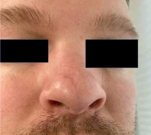
Figure 5: 3-months following complex repair with smooth contour. Scars may require surgical revision.
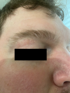
Figure 6: Well-healed upper eyelid laceration repair.
