Degloved Plantar Wound

Bruce A. Kraemer, M.D.
History:
Forty-five year old motorcyclist injured his left foot after he was side-swiped by a car and his left foot became entangled in the motorcycle and he sustained a complex plantar laceration with degloved flaps of the majority of the plantar surface.  He also sustained a right shoulder acromioclavicular separation. He was a non-smoker, had no significant medical problems and did not take any routine medications.

Findings:

.

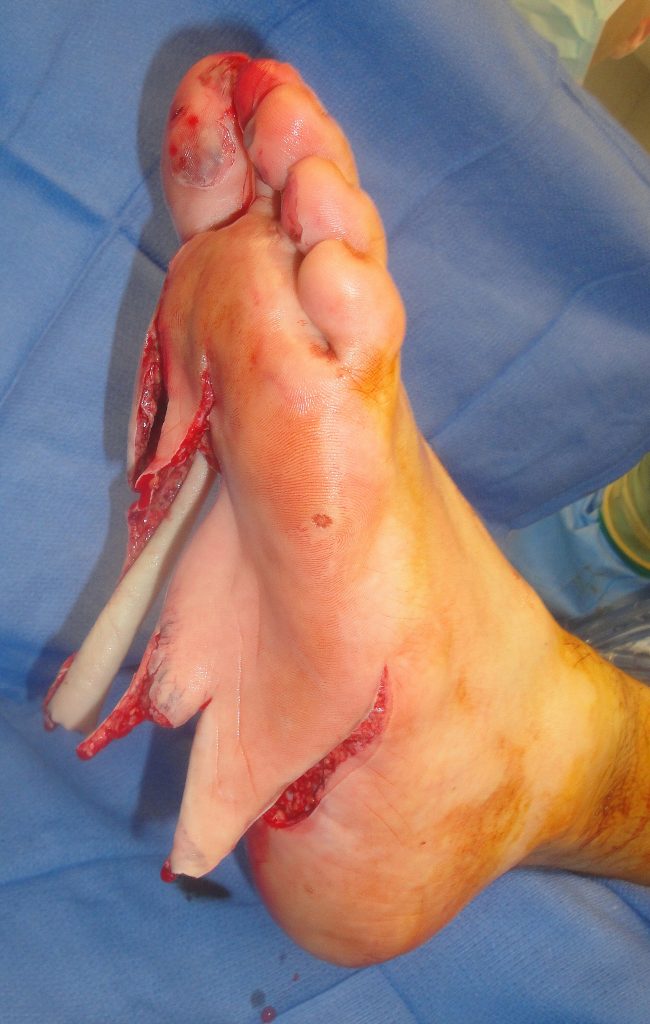
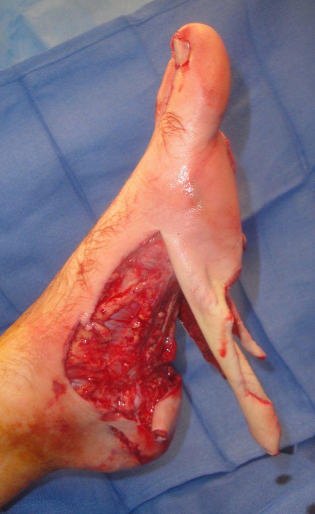
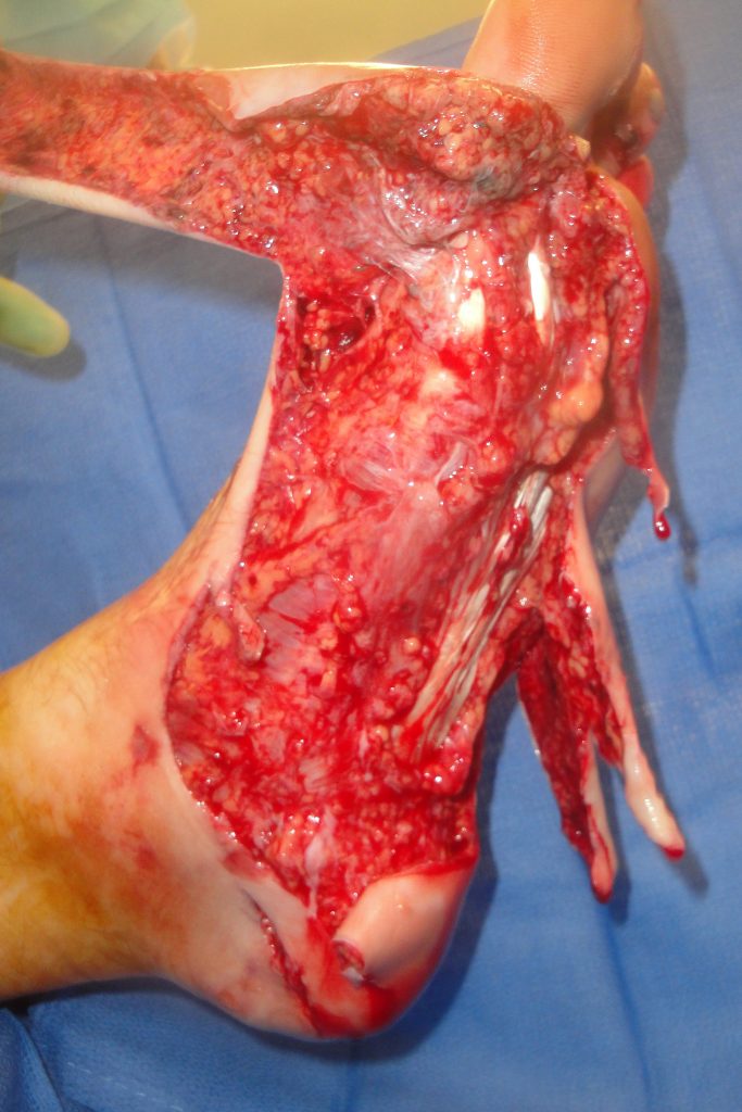
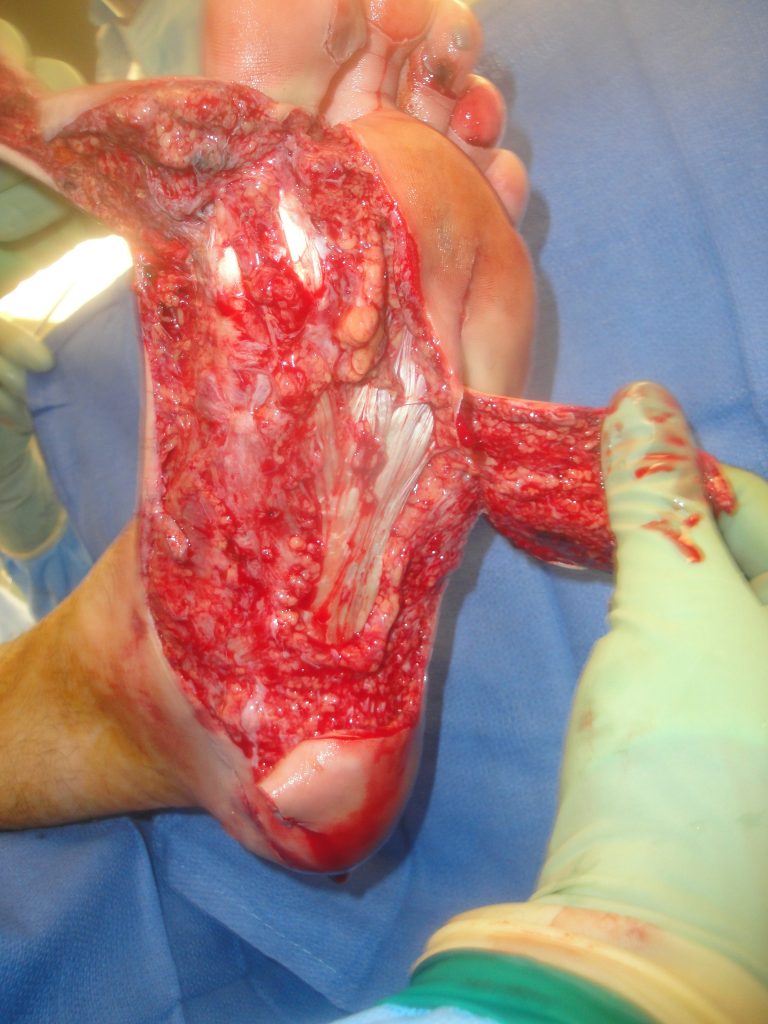
Fig.1. Complex plantar degloving lacerations of the left foot on the day of injury.

The plantar wounds involved a majority of the left foot plantar surface as shown

Diagnosis:

Complex degloving lacerations of a majority of left foot plantar surface without associated fracture.

Differential Diagnoses:

None- Acute Injury

Workup Required:

PA, lateral and oblique x-rays of the foot are needed to look for possible associated fractures, dislocations or retained foreign bodies.  Wound bed cultures should also be taken after initial debridement to assess the initial bacterial contamination of the wound which often proves useful should infection develop during the early phases of treatment.

Plan:

The treatment goal was to salvage as much of the plantar surface as possible and to provide a durably healed, stable, sensate plantar surface able to accommodate normal footwear.
The wound needed to be carefully debrided and then the areas of tissue deficit treated.  As it was difficult to acutely determine ultimate flap viability- especially with distally based flaps- it was decided to determine plantar flap viability after the patient was fully vascularly resuscitated and the wounds treated with an extracellular wound device and negative pressure wound therapy.

Expertise Needed:

A plastic surgeon with particular interest in soft tissue reconstruction should assess the patient and make the determinations as to viability and salvageability of the plantar tissues.  Knowledge of the use of Extracellular Matrix Wound Healing Devices is desired as these devices facilitate healing through a process termed “site-specific constructive remodeling”- promotion of the formation of more normal tissue which often minimizes scarring.  This M-2 macrophage directed healing is highly desirous in treating a serious plantar soft tissue injury.  Placing these devices in the wound and then surrounding them with tissues on all side provides the ideal environment for constructive remodeling.

Treatment:

Initial wound cleaning and debridement was accomplished with syringe and then judicious hydrosurgical dissection and debridement (Figure 1).  Minimal trimming of the distal ends of the flaps was done initially to let viability.be determined at a later date after full circulatory resuscitation and treatment with an Extracellular Matrix wound devices and negative pressure wound therapy had been attempted.

The wound be was treated with 2,500 mg of Urinary Bladder Matrix-Extracellular Matrix (UBM-ECM) powder and bilayer lyophilized UBM-ECM wound sheets (10 x 15 cm & 7 x 10 cm) (Figure 2).  The flaps were loosely tacked down (Figure 3) and then a dressing of vaseline impregnated gauze, black foam and a polyurethane sheet NPWT device was applied (Figure 4).  The NPWT was run at 80 mm Hg. on a continuous setting.

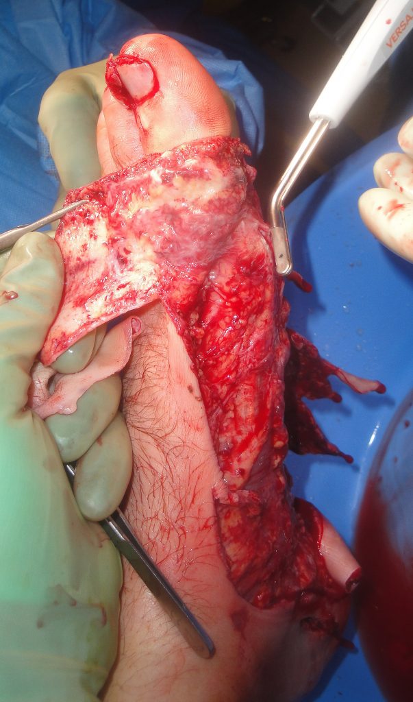
Fig.1. UBM treatment

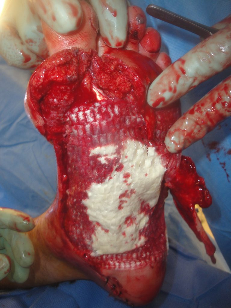
Fig.2. Further UBM treatment

Fig.3. Full plantar aspect after UBM treatment

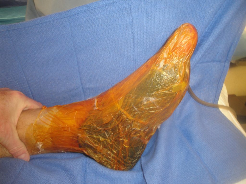
Fig.4. Negative pressure dressing over UBM.

Follow Up:

Negative pressure wound therapy was used for the first 3 weeks and then again for 1 week after split thickness skin grafting done at post-operative week 4.  During this time the NPWT dressing was changed weekly and the outer wound slough and desquamated skin was rinsed off before replacing a new vaseline impregnated gauze and piece of black foam.  Moisture retentive dressings were used until there was wound closure.  This consisted of either a polyurethane sheet dressing or hydrogel with a non-adherent  dressing.  He was able to ambulate small distances beginning at 3 months and was fully ambulatory with a padded sole shoe at 4 weeks.  He regained complete sensation of his plantar surface and was able to ambulate on 4-5 hour hikes and felt no serious limitations.  He had normal tissues over the main pressure points of his plantar surface and the healed incision lines had soft supple scars.

Clinical Note– Over time there was replacement of the skin grafted wound with normal plantar tissues which was not scar retraction of the skin grafts which can be seen in the clinical photos.

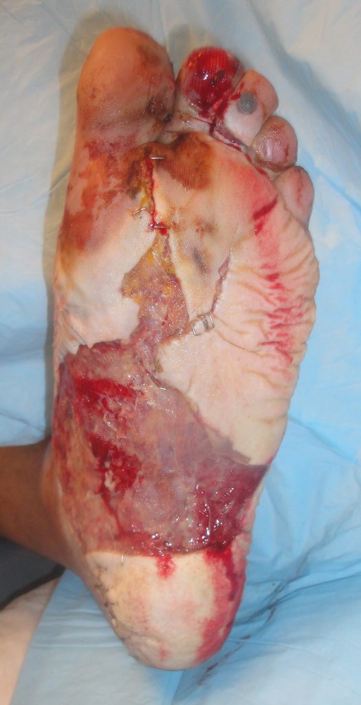
Fig.5. Post-OP healing week 2

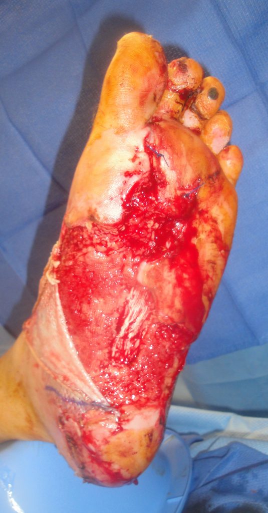
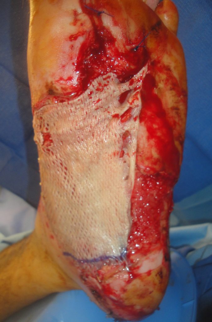
Fig.6. Wound healing at week 4 at the time of plantar split thickness skin grafting

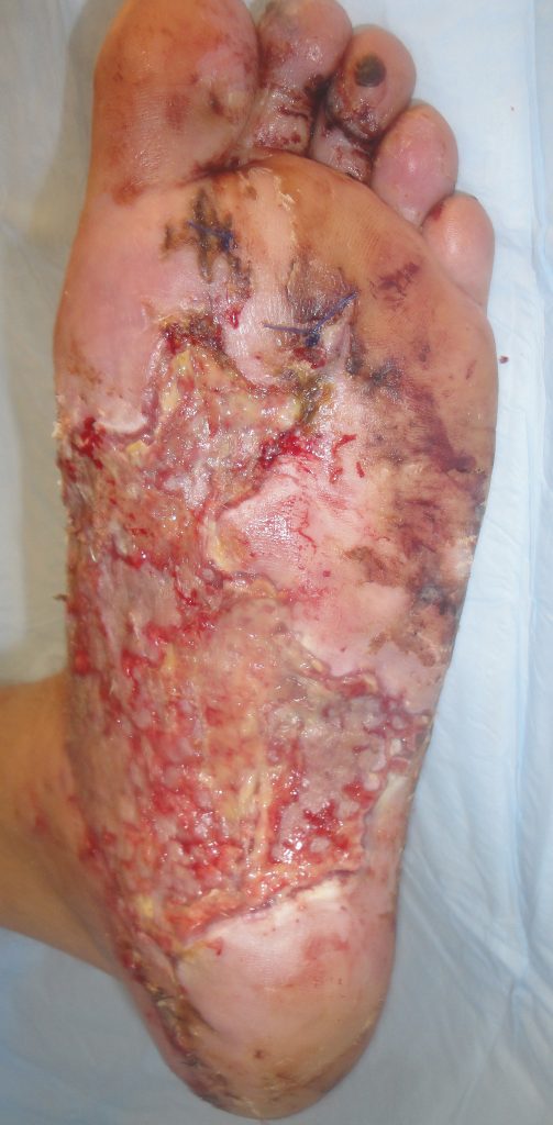
Fig.7. Plantar healing 2 weeks after split thickness skin grafting

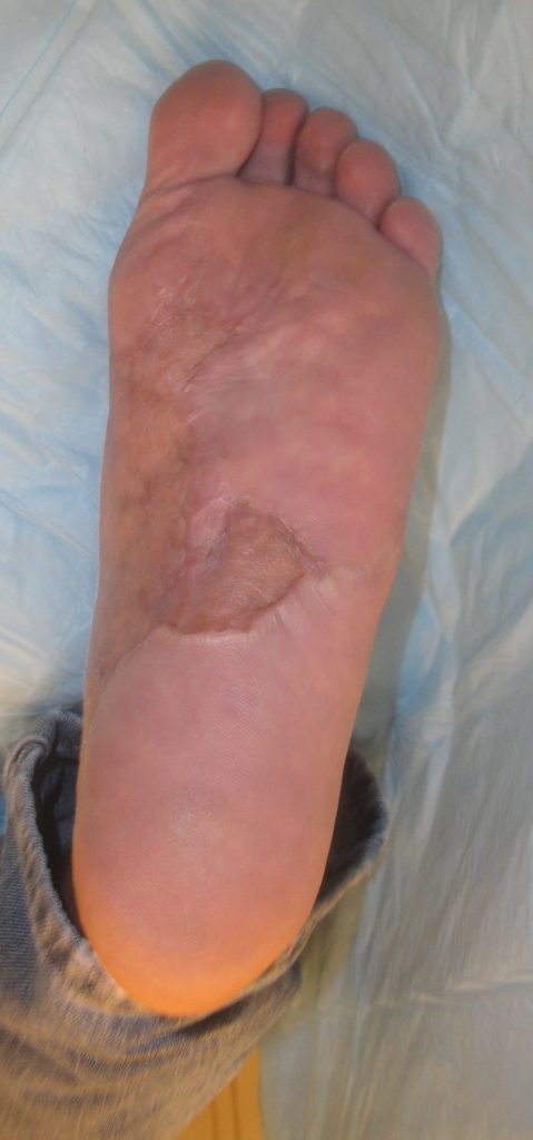
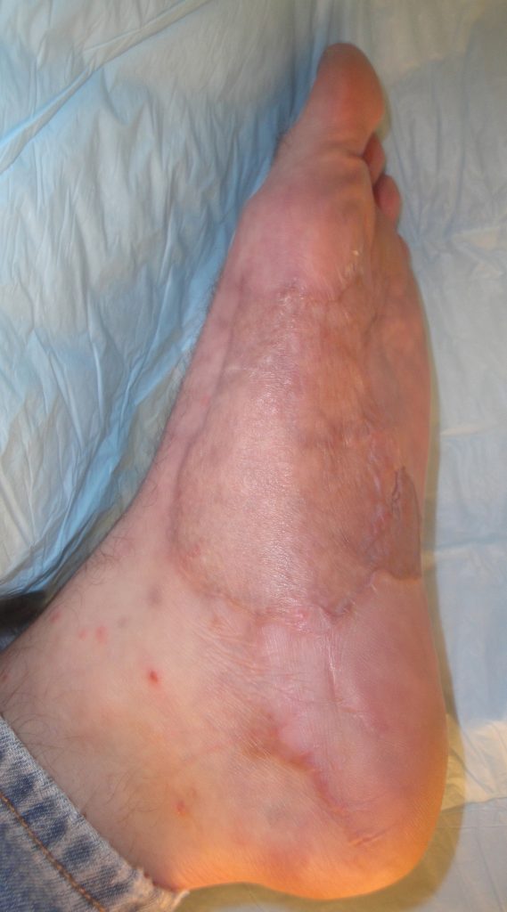
Fig. 8. Final plantar wound appearance 14 months post-injury

Print Friendly, PDF & Email

References

Geiger SE, Deigni OA, Watson JT, Kraemer BA. Management of Open Distal Lower Extremity Wounds With Exposed Tendons Using Porcine Urinary Bladder Matrix. Wounds- A Compendium of Clinical Research & Practice 2016;28(9):308-316.
Kraemer BA, Geiger SE, Deigni OA, Watson JT. Management of Open Distal Lower Extremity Wounds with Concomitant Fracture Using Porcine Urinary Bladder Matrix. Wounds- A Compendium of Clinical Research & Practice 2016; 28(11):387-394.
Kraemer BA: Management of Complex Distal Lower Extremity Wounds Using a Porcine Urinary Bladder Matrix (UBM-ECM). In: Shiffman MA, Low M Eds. Plastic and Thoracic Surgery, Orthopedics and Ophthalmology. Cham, Switzerland: Springer International Publishing AG; 2020: 3-29.
https://pubmed.ncbi.nlm.nih.gov/27701126/
https://pubmed.ncbi.nlm.nih.gov/27861131/
https://link.springer.com/book/10.1007/978-3-030-10710-9

Related Articles