History:
70-year female with a past medical history of stage IV spindle cell sarcoma of the left posterior upper extremity that was resected in 2014 and treated with adjuvant radiation. She developed a local recurrence requiring repeat excision and radiotherapy in 2019 which was complicated by a radionecrotic wound with a chronic draining sinus tract from a seroma cavity. She was first seen in the plastic and reconstructive surgery clinic in February of 2020, and after failing a trial of local wound care/compression, she underwent radical excision of the wound and reconstruction with an extended left pedicled latissimus dorsi myocutaneous flap.
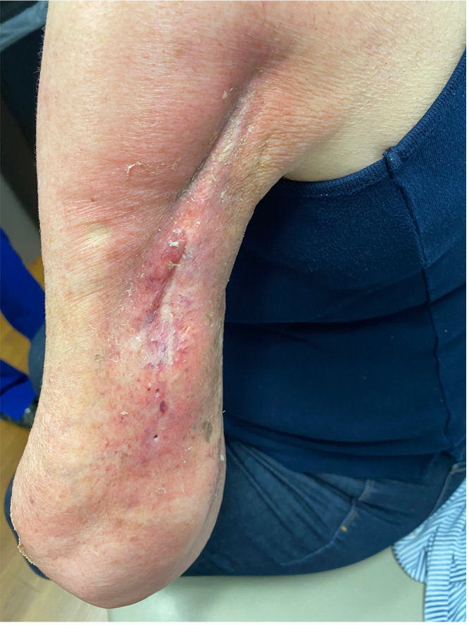
Fig. 1. Preop photo showing wound with pinpoint sinus tract
Findings:
Resection of radiated necrotic tissue extending into a cavity overlying the humerus resulting in a 20 x 10 cm defect with exposed bone and vasculature
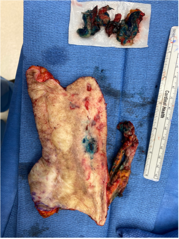
Fig.2. Wound/seroma cavity excised en bloc; photo showing methylene blue dye used to assist dissection
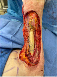
Fig.3. Resultant defect showing exposed humerus
Treatment:
The patient was taken to the operating room and placed under general anesthesia. She was positioned with the left side up using a bean bag for adequate exposure of the donor site and the left upper extremity. Methylene blue was injected into the chronic draining sinus to delineate the involved tissue and the radiated tissue and seroma cavity were sharply excised en block down to the humerus. The bone was then curetted, and the wound was irrigated with antibiotic solution. A template of the defect was made and transposed over the left latissimus for flap planning. The flap needed to be 10 cm wide and 30 cm in length and to account for the 180-degree turn and these incisions were marked out.
The markings were then incised through the skin and subcutaneous tissue down to the latissimus muscle and the surrounding tissues were mobilized to expose the borders of the muscle and assist with closure. The posterior border of the muscle was transected, and the anterior border was elevated off the underlying serratus anterior. The distal aspect of the muscle was then transected, and the flap was elevated from inferior to superior to develop this as a superiorly based flap off the thoracodorsal vessels. After full elevation of the flap and protection of the pedicle, it was rotated 180 degrees to fill the defect. A small skin bridge separated the defect from the donor site and this area was excised as it had been radiated as well. Two drains were placed, one in the defect and one in the donor site, and the flap was inset with 3-0 Monocryl and a running Prolene suture. The donor site was closed with 2-0 vicryl in a figure of eight fashion, utilizing penetrating towel clips for tissue creep and approximation, and the final layer was closed with a 3-0 V-loc.
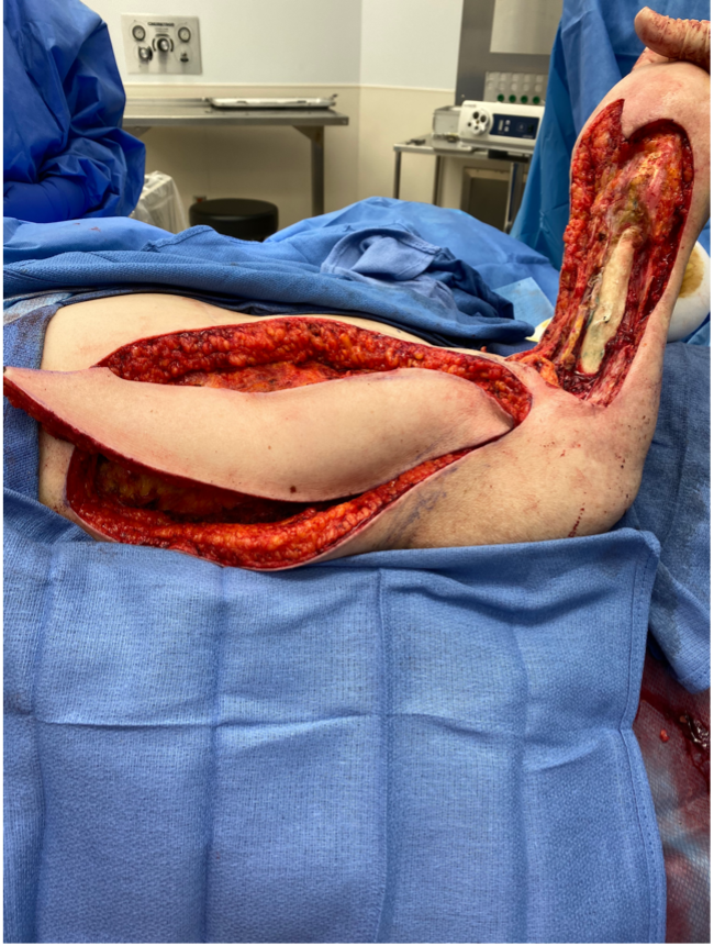
Fig.4. Elevated latissimus dorsi myocutaneous flap
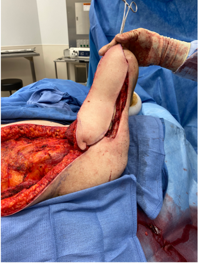
Fig.5. Latissimus dorsi flap rotated into defect
Follow Up:
The patient was seen one week after surgery and both drains were removed. Sutures were removed at three weeks and there were no signs of wound breakdown or dehiscence. She was again seen at three months and doing well from a surgical standpoint.
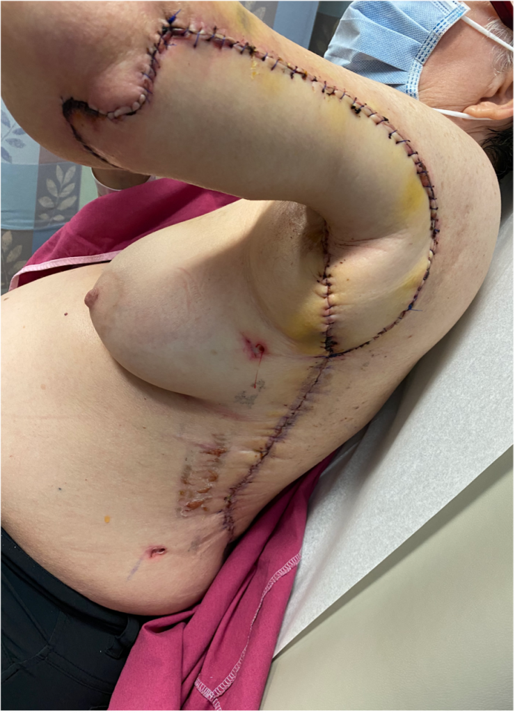
Fig.6. 1 week post op
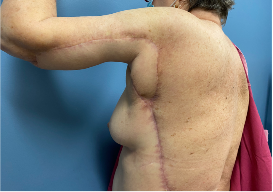
Fig.7. 3 months post op
