Findings:
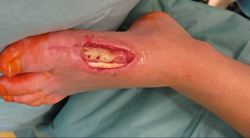
During the intraoperative examination in the department of plastic and reconstructive surgery, we measured a soft tissue defect of about 8 x 4 cm on the arch of the left foot. The musculus flexor hallucis longus tendon and the first metatarsal bone were exposed.
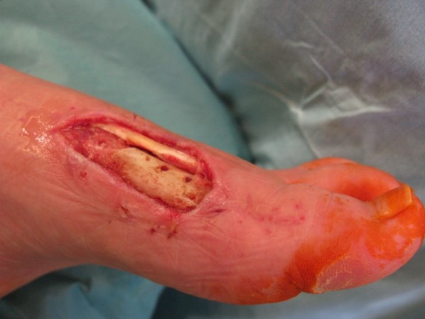
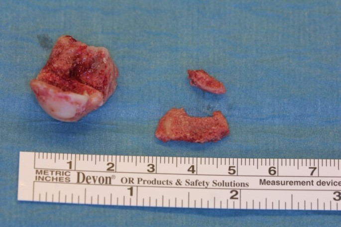
The caput of os metatarsale I was nonviable and therefore needed to be removed.
Treatment:
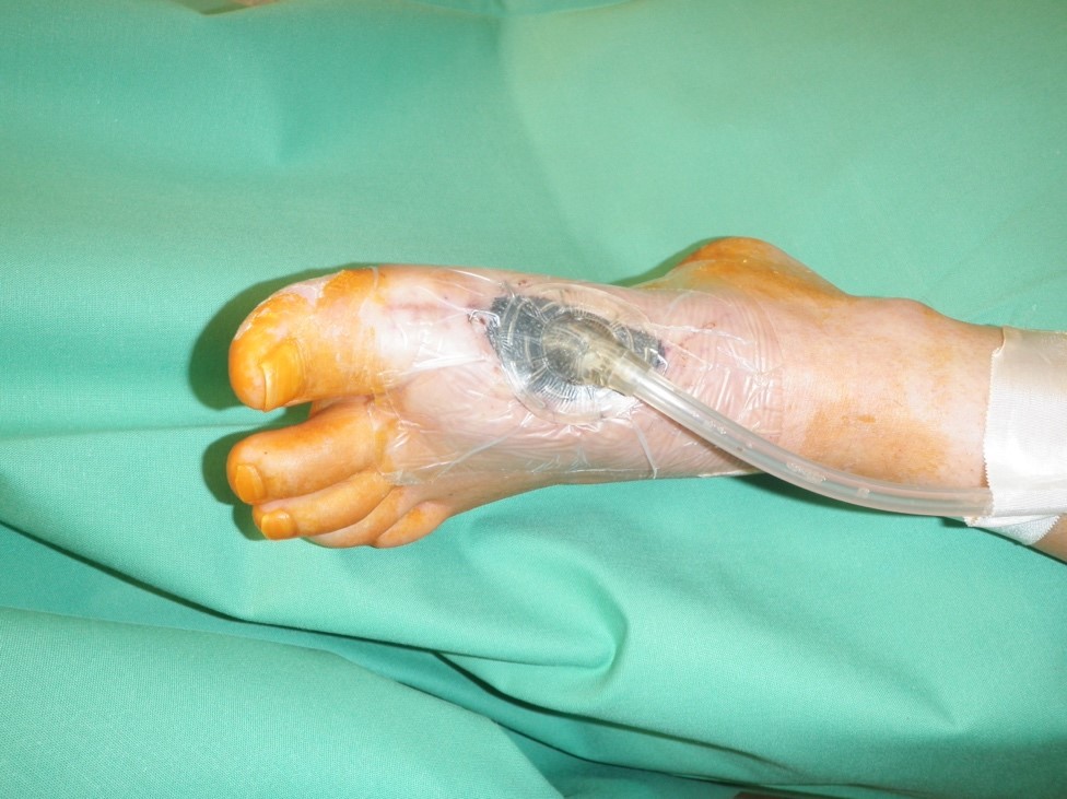
The patient had multiple VAC-therapy cycles for wound conditioning.
After the debridement, the entire soft tissue defect of the foot with exposed tendons and bone were visible.
The caput of os metatarsale I was nonviable and therefore needed to be removed.
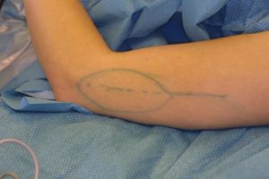
The plastic surgeon decided to perform a lateral upper arm- free flap. Firstly, the vessels at the recipient-site were prepared. Arteria dorsalis pedis and two accompanying veins were identified. Subsequently, the lateral upper arm- free flap was dissected.
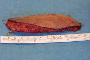
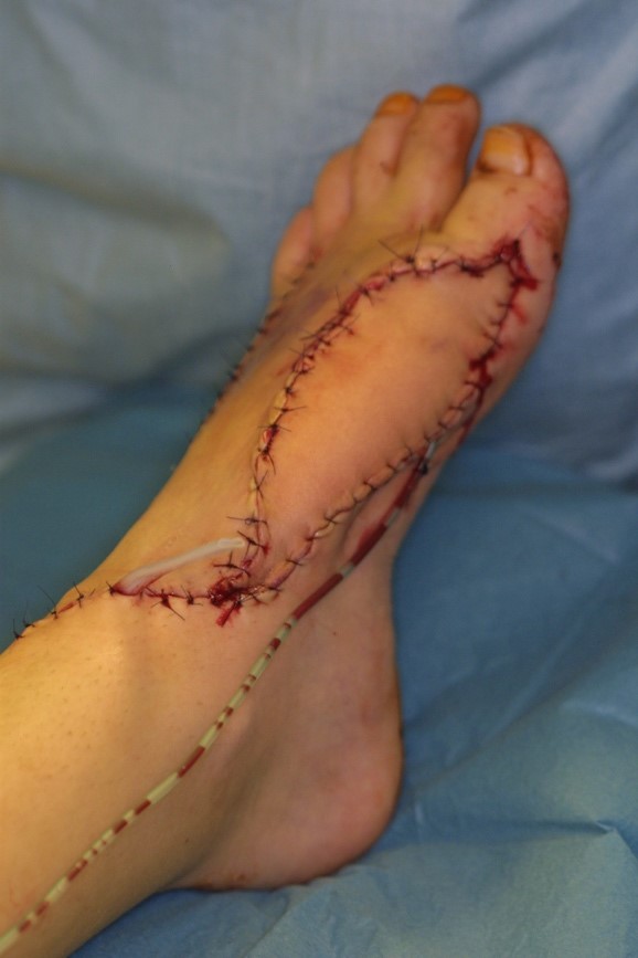
Using an operating microscope, the free flap was anastomosed end-to-end to the Arteria dorsalis pedis. Furthermore, two veins were anastomosed to ensure a sufficient venous backflow. Drains were placed and subcutaneous and skin sutures were performed.
Follow Up:
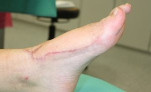
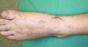
The patient was observed at the intensive care unit for one night after surgery. The free-flap was monitored hourly in the first 24 hours by a clinical examination and doppler ultrasonography. For the next 24 hours, the free flap was monitored every two hours and the next day every four hours. The drains were removed after a few days. The sutures were all removed in post-operative week 3. The patient had physiotherapy. The patient had to take Aspirin for 10 days and got a thrombosis prophylaxis and a sufficient pain medication. After patient discharge the patient had control examinations in our clinic. The wound healing was without complications. The patient was satisfied as she had normal gait in normal shoes and was not compromised in her daily activities.