Chronic Venous Stasis Ulcer

H. Dan Derbes, MD
, Rummana Aslam, MD

History:

An 84 Year-old-male presented to our Wound Care Clinic with a history of a non-healing ulcer on his right lower extremity which had been present for approximately 1 year. His ulcer was associated with significant discomfort in and around the ulcer bed, as well as moderate bilateral lower extremity edema. He was capable of independent ambulation at baseline, and did not recall an inciting incident for how he acquired the wound. His medical history was significant for Hypertension, Hyperlipidemia, Atrial Fibrillation on Eliquis, Esophageal Cancer, Deep Vein Thrombosis with Pulmonary Embolism, Gout, Chronic Kidney Disease Stage 3, and Diabetes Mellitus with bilateral lower extremity neuropathy. He had also been counseled in the past that he had venous insufficiency of the bilateral lower extremities, and had been using custom fit compression garments on his left lower extremity (LLE), but had not been able to apply his own compression on the right lower extremity (RLE) due to his ulcer. He had been receiving compression bandaging from his home health nurses three times weekly for his right lower extremity without ulcer improvement. Approximately 6 months prior to presentation to our clinic, he had undergone an endo-venous ablation of the Right Greater Saphenous Vein (R-GSV), but this did not improve his edema or wound. Surgical history is significant for a bio-prosthetic Aortic Valve Replacement.

Findings:

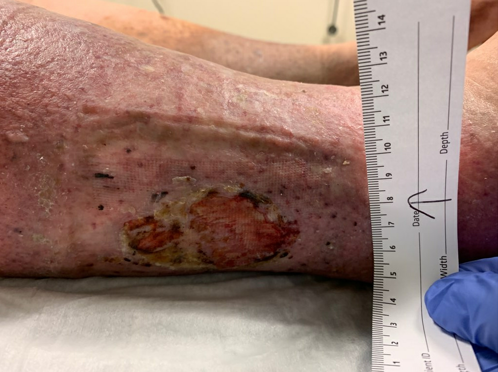
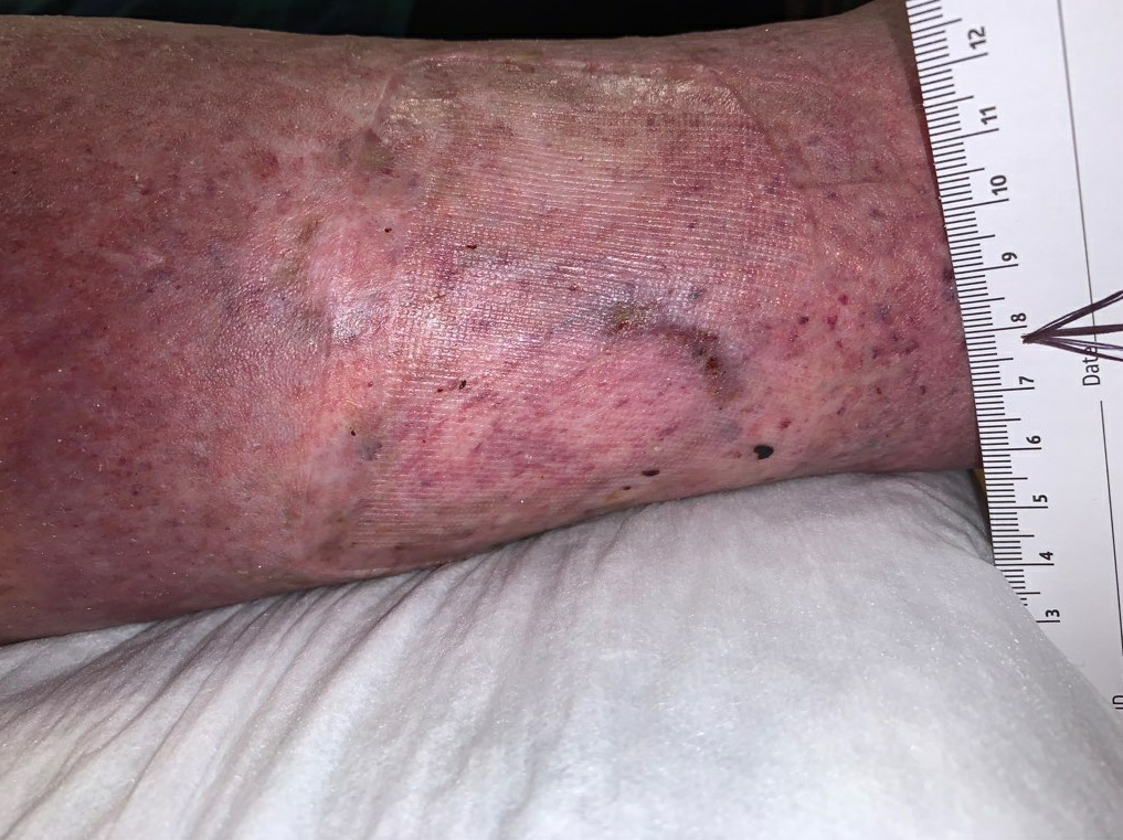
At our first encounter, he was noted to have an ulcer on the lateral aspect of his RLE, about 6cm proximal to the lateral malleolus. The ulcer measured 6×4.3cm (25.8cm2), and had indurated, rolled edges, with moderate serous drainage. The ulcer was not foul smelling, and there was only scant fibrinous exudate in the ulcer bed. Due to discomfort, we elected not to debride it at the first visit. In further discussion with him, we also noted that when he sat in his recliner in the evenings, there was an area of pressure and friction caused by the footrest on his recliner which corresponded directly to the position of the ulcer. He was advised to put a pillow under his feet, to minimize the direct pressure on the ulcer by the foot rest.

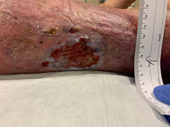
RLE Venous Stasis Ulcer at presentation to our Wound Care Center. Size: 25.8cm2


Diagnosis:

Bilateral Lower Extremity Venous Insufficiency with Venous Hypertension
Venous Stasis Dermatitis with Hemosiderosis
Right Lower Extremity Venous Stasis Ulcer (chronic)

Differential Diagnoses:

This patient clearly had a venous stasis ulcer, but it was important to determine whether there were other co-morbidities that were also contributing to his chronic inability to heal the wound, such as poorly controlled Diabetes Mellitus, Peripheral Arterial Disease (PAD), or chronic wound infection. There was also consideration for unresolved venous insufficiency that might be amenable to further lower extremity vascular procedures, such as a repeat ablation of a recurrent GSV insufficiency due to GSV recanalization, or unresolved perforator vein insufficiency which might be amenable to perforator vein sclerotherapy.

Workup Required:

To rule out concurrent lower leg pathology, complete Ankle Brachial Indexes and Toe Brachial Indexes with capillary wedge pressure testing and peripheral vascular resistance testing was performed bilaterally, and was unremarkable.
The patient also underwent full doppler venous ultrasound of his bilateral lower extremities, and mild reflux was noted bilaterally in the femoral and popliteal veins, but his R-GSV was still occluded, and there was no evidence of active DVT. There was no evidence of perforator venous insufficiency. We concluded that there was no significant opportunity to optimize his venous insufficiency through further ablative procedures.
Wound cultures were performed, and grew 3+ Corynebacterium striatum and 3+ Staphylococcus aureus. Given that the patient had no leukocytosis, was afebrile, and had no clinical signs of local infection, this was determined to be a chronically colonized wound, rather than an acute/chronic wound infection. We determined that systemic antibiotics would not be of benefit.

Plan:

The primary treatment for venous stasis ulcers, compression therapy, was initiated to help treat both the lower extremity edema as well as the venous insufficiency. We applied an ulcer dressing consisting of cadexomer iodine, followed by an absorptive foam layer, followed by a multilayer compression bandage (Coflex lite).
We also obtained wound cultures, which ultimately grew 3+ Corynebacterium striatum and 3+ Staphylococcus aureus. We assumed that the wound was colonized even prior to these results, which was why we chose cadexomer iodine as the base layer of his dressing.
We counseled him to use a pillow to pad his legs in the evenings when he sat in his recliner, as his recliner’s foot rest was old and poorly padded, and causing pressure on the ulcer.
We also encouraged him to continue his regular follow up with his endocrinologist regarding his Diabetes.
We educated him on the importance of compression therapy for treating his ulcer and preventing it’s recurrence once it eventually healed. This teaching was reinforced at every visit.
We asked that he discontinue his 3x per week VNA visits, so that all wound care be performed by the wound care physician and our staff. This allowed for closer monitoring of the status of the wound, and its response to our treatment and any treatment changes, it also improved the consistency of his care, since all care would be delivered by a single team.
Follow up was set at 1-2 times per week as possible.

Expertise Needed:

Vascular surgery evaluation had already been performed, and a previous R-GSV ablation had also been performed.
Wound Care Physician took charge of the wound therapy plan, and this plan was executed by the Wound Care Nurses at our clinic.
VNA wound care nurses were discontinued to prevent overlapping or conflicting care.
Endocrinology and Primary Care Provider for discussion about diabetes management, though no significant changes were made to his regimen during this time.

Treatment:

Compression was initiated with Coflex lite multilayer bandage over an absorptive foam, over a base layer of cadexomer iodine.
Compression: Coflex lite included an absorptive foam layer, which was helpful at managing mild levels of wound exudate, while still providing adequate compression. We like this dressing system for its combination of compression and absorptance. If Coflex had produced too much compression for comfort, we would have used a lighter compression sleeve, such as SpandaGrip.
Base dressing layer:
Cadexomer Iodone was chosen initially due to its ability to help absorb exudate, which is helpful early in the treatment of venous ulcers. It is also a topical antiseptic, effective in addressing bacterial colonization with or without biofilm. Once the volume of exudate had diminished, and the wound edges became dried instead of macerated, we switched to topical bactroban, to help keep the ulcer appropriately moist while still providing coverage against colonization which had previously been noted. Once we were confident that the colonization issue had been adequately addressed, we to switched to enzymatic debridement with Santyl. In a situation where both fibrinous exudate and colonization were noted, we addressed the colonization first. We have found Santyl helpful at degrading and clearing fibrinous slough in between sharp debridements. Once bacterial colonization had been addressed, slough was addressed with Santyl to further aide in wound healing.

Follow Up:

At our first follow up with him 1 week later, he was tolerating his compression dressing (which had been in place for 7 days), and the ulcer surface area had improved to 18.5cm2, with a healthy bed of beefy red granulation tissue forming. He still had moderate edema, and his wound was tender to palpation so we again deferred debridement until his discomfort was under better control.

1 week follow up, healthier base with beefy red granulation tissue. Edges are still indurated and rolled. Surface Area 18.5cm2

At our 2 week follow up his pain had improved significantly, but he did complain of discomfort in the first couple of days after the new compression had been applied last time. His pain improved gradually throughout the week, but he was apprehensive about receiving the same level of compression again. At this time we elected to perform a debridement of the edge of the wound, as it was still indurated with rolled edges. We performed an excisional debridement using a sharp curette, only resecting non-viable tissue and slough, and freshening the edge of the ulcer until a slight ooze of blood was encountered. Hemostasis was achieved with gentle pressure. A similar debridement was performed on week 3. Between the debridements during the second and third weeks, the ulcer had been resected back to its original size, but this time with significantly healthier edges and a cleaner ulcer base with healthy granulation tissue in the ulcer bed. Due to his apprehension with a high level of compression, the compression level was adjusted to his comfort using a Spandagrip sleeve, a more mild form of compression. We would have preferred higher levels of compression, if the patient had tolerated this.

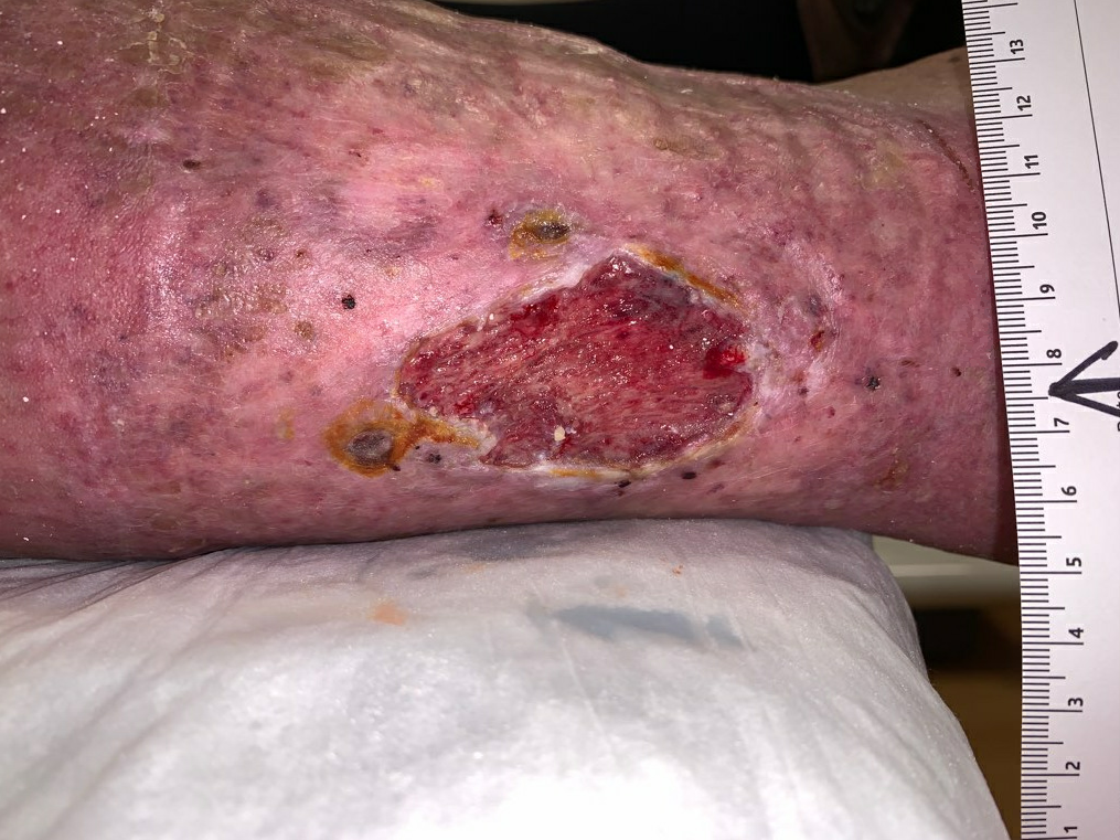
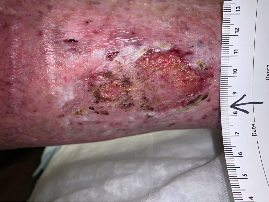
Week 3: Edges are flatter and less rolled, healthy granulation tissue in ulcer bed. Size: 24.3cm2


Week 3.5, wound edges are dry and non-viable, thin exudate, gentle debridement will be performed

Week 3.5 post debridement, the edges were freshened slightly, and some scant slough was removed

By week 4 he had evidence of epithelialization over approximately 70% of the initial wound bed, and since he had been compliant with his compression therapy, his edema was also markedly improved. As his edema improved, his exudate had significantly decreased, resulting in drier wound edges, rather than the macerated, rolled edges that he started with. To adapt to this, we changed his base layer in his dressing from cadexomer iodine (absorptive) to bactroban ointment (helps to maintain moisture).

Week 4: Significant progress in re-epithelialization, edges dry and flat

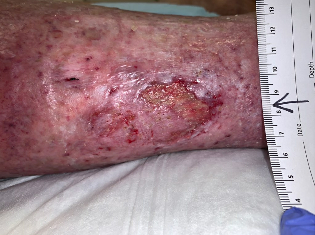
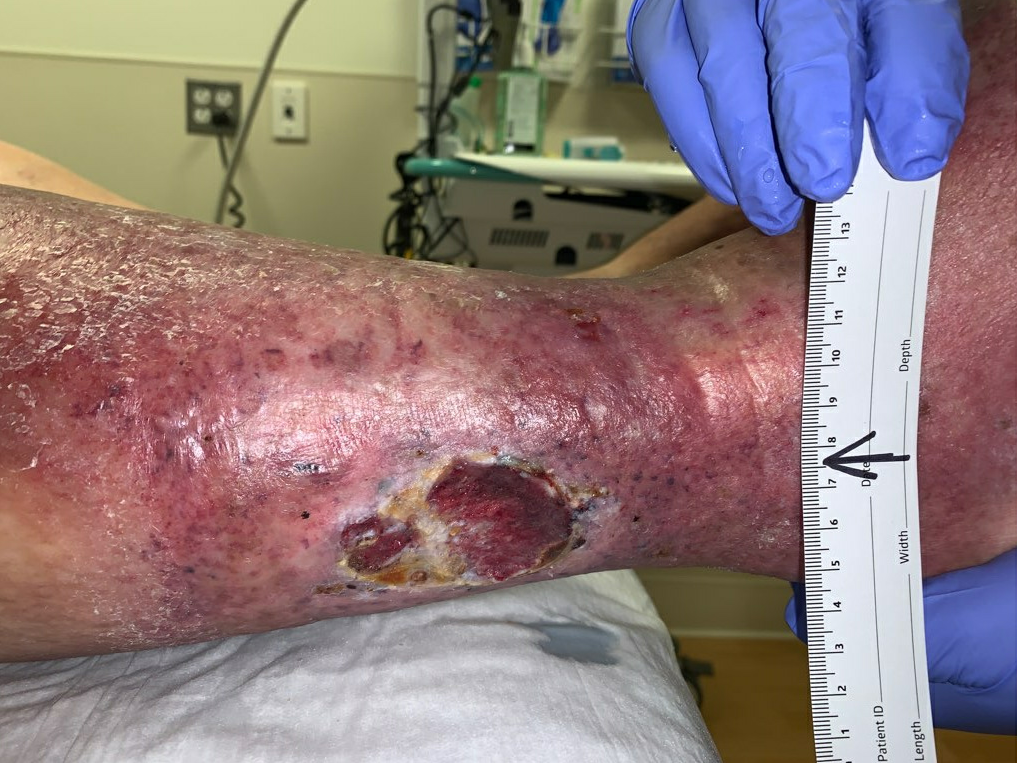
Healthy granulation tissue noted under dried skin edges/eschar after debridement. Size: ~9cm2 open areas


By week 5 we were able to get him a pneumatic compression pump to use at home for 1 hour per day in addition to his compression bandages. He had initially been given 30-40 mmHg calf high compression stockings for use on his left leg only, as his right leg was receiving compression from our wound dressing. He had been compliant with using this compression, but when questioned about willingness to continue long term use, he confessed that he would not be willing to continue using this level of compression indefinitely, as it was uncomfortable during the day. To improve his likelihood of compliance, we changed his compression prescription to 20-30 mmHg compression stockings, which he found significantly more comfortable, and he stated that he was willing to use these indefinitely, as we had continued to reinforce the importance in maintaining compression therapy forever, as this was the best way to prevent recurrence of his chronic stasis ulcer. He also agreed to continue using the compression pump nightly. He continued following up 1-2 times per week, and his wound received more gentle, serial debridements for the purpose of removing slough and freshening the edge of the ulcer.

By the 6th follow up week, he had made significant improvement in the size of the ulcer. He also denied any significant discomfort, both at baseline and with wound care and applying compression.

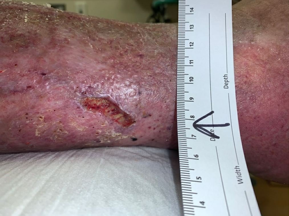
Ulcer was significantly smaller with healthy epithelium covering most of the ulcer bed. Size: 3.5cm2

As his pain had improved significantly, we resumed Coflex Lite as his primary form of compression. We also switched his Bactroban Ointment for Santyl, to allow for enzymatic debridement of the minimal slough that was still present in the wound. While we did not repeat a culture of the ulcer bed, we believed that the colonization had been adequately addressed by this time. His follow up by week 8 demonstrated nearly complete healing, with less than 3cm2 of surface area left, and evidence of new epithelialization over most of the remaining area.

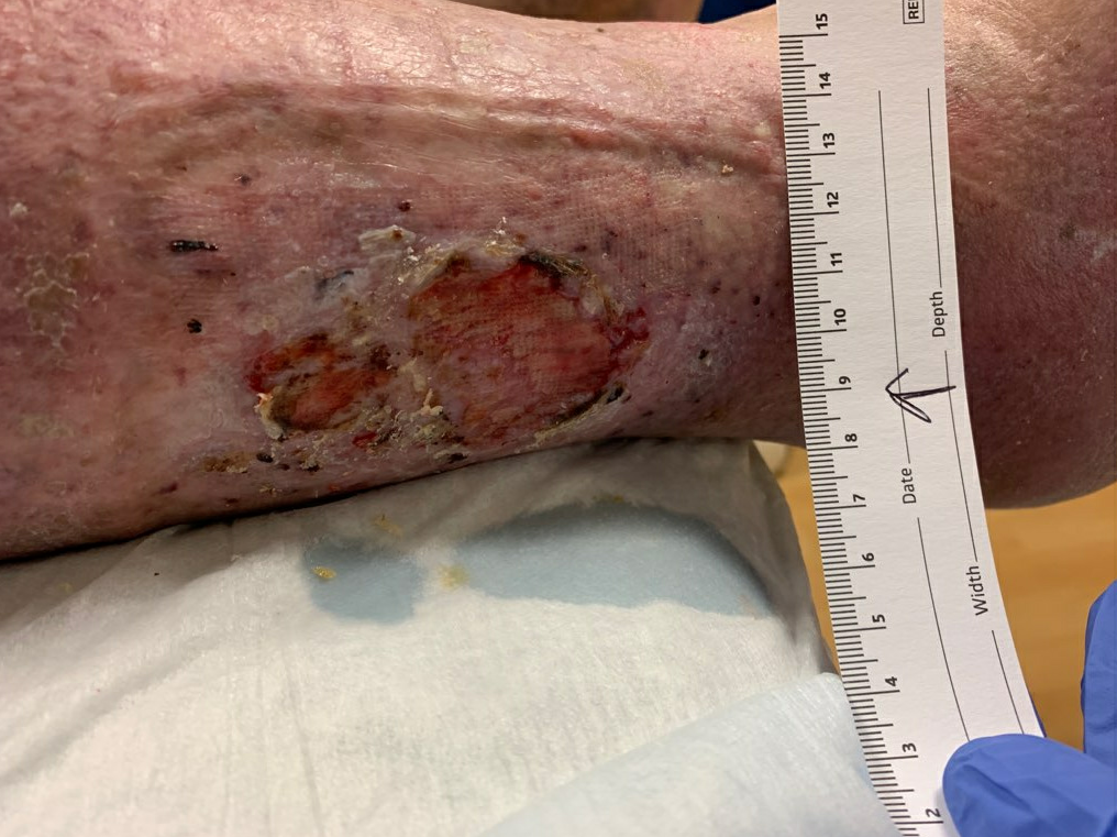
Week 8: Ulcer bed is healthy, there are small islands of epithelium scattered among the remaining open area. Size: <3cm2

His ulcer was noted to have fully re-epithelialized at his 9 week follow up. His edema had markedly improved, there was only trace edema remaining. He was counseled at this time to maintain his compression therapy with stockings (20-30 mmHg calf high) forever, as this is the single best way to prevent recurrence. He should wear his stockings during the day (~12 hours at least), and remove them at night when he was able to sit and elevate his feet. He was advised that physical activity was also helpful in preventing edema, in combination with compression therapy. He was instructed to return early if another wound developed, as he remains at high risk for developing future chronic wounds.

9 Weeks: Full Re-epithelialization

Print Friendly, PDF & Email

References

Bush RG, Bush P, Flanagan J, et al. Factors associated with recurrence of varicose veins after thermal ablation: results of the recurrent veins after thermal ablation study. Scientific World Journal. 2014;2014:505843. Published 2014 Jan 27. doi:10.1155/2014/505843
Marston W, Tang J, Kirsner RS, Ennis W. Wound Healing Society 2015 update on guidelines for venous ulcers. Wound Repair Regen. 2016 Jan-Feb;24(1):136-44. doi: 10.1111/wrr.12394. PMID: 26663616.
Partsch H, Mortimer P. Compression for leg wounds. Br J Dermatol. 2015 Aug;173(2):359-69. doi: 10.1111/bjd.13851. Epub 2015 Jun 12. PMID: 26094638.
https://www.ncbi.nlm.nih.gov/pmc/articles/PMC3921954/
https://onlinelibrary.wiley.com/doi/epdf/10.1111/wrr.12394
https://onlinelibrary.wiley.com/doi/10.1111/bjd.13851

Related Articles