Chin laceration

Olivier F. Noel, M.D., Ph.D.
, Jackie Oh, M.D.
, Omar Allam, M.D.

History:

A 26 year old male with left chin laceration sustained during a motor vehicle accident. He denied any loss of consciousness or any other injuries. He reported moderate bleeding from his left chin that was controlled with pressure. TDAP was given in the ED. No further imaging or labs were obtained by the emergency department. The Plastic and Reconstructive Surgery team was consulted for the chin laceration.

Findings:

On exam, 4 cm long,  deep laceration to the left chin, with exposed mentalis muscle. The laceration was deep but with no intraoral component. There was no loss of teeth or evidence of facial fractures.

Figure 1-2. Show left chin laceration.

Diagnosis:

Complex laceration of left chin.

Differential Diagnoses:

Chin laceration, r/o injury to mental nerve.

Workup Required:

Cranial Nerves 2-11 were tested and were without deficit although there were mildly decreased sensation to the left chin compared to the right. The laceration was inferior to the mental foramen and the mental nerve was intact. No intraoral lacerations, malocclusion, or loose teeth were noted on exam. Consent was then obtained for irrigation, debridement, and closure of his chin laceration.

Plan:

Repair of the laceration under local anesthesia in the Emergency Department.

Expertise Needed:

Plastic surgeon.

Treatment:

The patient was placed in the supine position with good lighting. The wound was copiously irrigated with saline and betadine solution. In the muscle, 4-0 Monocryl sutures were used. In the superficial layer, 6-0 Monocryl dermal sutures were used to approximate the dermal edges. 6-0 Prolene interrupted sutures were used for the best skin approximation. Bacitracin was applied to the wound and the patient was instructed to apply Bacitracin ointment twice daily until follow up. He was also instructed to avoid sun exposure and use SPF-30 or higher lotion if exposed to sun for the next year. This was done in order to decrease the risk of hyper pigmentation of her scar. He was scheduled for follow up in 1 week for suture removal. He received 5 days of prophylactic antibiotics. At the follow up visit, his wound was healing well, the sutures were removed, and the patient was instructed to apply Vaseline to the healed laceration until there was no more crusting.

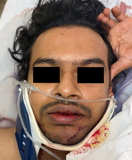
Figure 3-4. Show repaired left chin laceration.

Follow Up:

One week for removal of non-resorbable sutures, followed by 1 month follow up for healing. Additional follow ups as needed depending on scar maturation and healing.

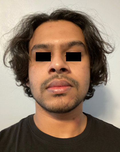
Figure 4. One month follow up.

Print Friendly, PDF & Email

References

Brown DJ, Jaffe JE, Henson JK. Advanced laceration management. Emerg Med Clin North Am. 2007 Feb;25(1):83-99. doi: 10.1016/j.emc.2006.11.001. PMID: 17400074.
Bhattacharya V. Management of soft tissue wounds of the face. Indian J Plast Surg. 2012 Sep;45(3):436-43. doi: 10.4103/0970-0358.105936. PMID: 23450264; PMCID: PMC3580340.
Franz MG, Robson MC, Steed DL, Barbul A, Brem H, Cooper DM, Leaper D, Milner SM, Payne WG, Wachtel TL, Wiersema-Bryant L; Wound Healing Society. Guidelines to aid healing of acute wounds by decreasing impediments of healing. Wound Repair Regen. 2008 Nov-Dec;16(6):723-48. doi: 10.1111/j.1524-475X.2008.00427.x. PMID: 19128244.
https://pubmed.ncbi.nlm.nih.gov/17400074/
https://www.ncbi.nlm.nih.gov/pmc/articles/PMC3580340/
https://pubmed.ncbi.nlm.nih.gov/19128244/

Related Articles