Juan Jose Lizardi, MD

History:

Patient is a 36 year old male with a mast medical history of hypertension, ICD placement, Aortic Valve replacement on warfarin who presents to the emergency department after being stabbed in the lateral left thigh leading to hematoma formation in the setting of a Profunda Artery branch extravasation and subsequent anterior thigh compartment release with complicated by large lateral thigh wound.

Findings:

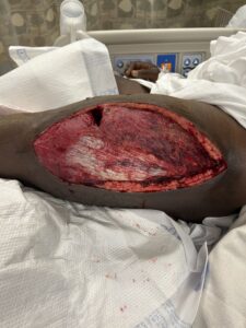
At Initial Evaluation: Large full thickness lateral thigh wound with exposed fascia and muscle. Overall healthy appearing wound bed with healthy bleeding from skin edges.

Diagnosis:

Large full thickness lateral thigh wound after fasciotomy in the setting of hematoma for patient on coagulation after suffering a stab wound to the thigh.

Workup Required:

Discuss case with Vascular Surgery colleagues to ensue that there are no further plans for surgical interventions from their stand point.
Routine pre-operative planning such also be undertaken including the following:
– Medical Co-morbidit optimization
– Nutrition Optimization (Consider albumin, pre-albumin, and nutrition consult if las are abberant)
– Type and Screen
– CBC
– BMP

Plan:

Operative intervention for coverage or large wound. The proximal and distal edges of the wound can be approximated with sutures. Careful attention not to try closing the wound at points with excessive tension. This will reduce the total surface area that need be covered with skin graft. For the remaining defect, a split thickness skin graft (harvested from contralateral thigh) was applied and sutured in placed to the wound. The split thickness skin graft was then covered and bolstered by a negative pressure wound therapy device (AKA a wound VAC) and kept in place for 5 days post-operatively.

Expertise Needed:

Though not a skill reserved for plastic and reconstructive surgeons, the adequate harvesting of a split thickness skin graft and its appropriate application is needed for coverage of this wound.

Treatment:

Underwent coverage of left lateral thigh wound with split thickness skin graft
Evaluation of Wound on POD5 after removal of negative pressure wound therapy device: Overall there appears to be good adherence of the split thickness skin graft. There is some underlying sanguineous output likely secondary to the patient’ anti-coagulative status

Follow Up:

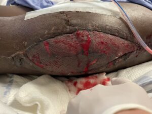
Patient was eventually discharged and then seen in clinic one month post-operatively where the following photo was taken. Overall the wound was healing well, with excellent take of the split thickness skin graft. Two site of granulation tissue near the mid-line superior aspect of the surgical site can be appreciated. Patient was counseled on best wound care practices and given return precautions.
Print Friendly, PDF & Email

References

Alkhalifah MK, Almutairi FSH. Optimising Wound Closure Following a Fasciotomy: A narrative review. Sultan Qaboos Univ Med J. 2019 Aug;19(3):e192-e200. doi: 10.18295/squmj.2019.19.03.004. Epub 2019 Nov 5. PMID: 31728216; PMCID: PMC6839671.
Walters ET, Kim KG, Dekker PK, Stimac GP, Mehra S, Elmarsafi T, Steinberg JS, Attinger CE, Kim PJ, Evans KK. Neither Antiplatelet nor Anticoagulant Therapy Increases Graft Failure after Split-thickness Skin Grafting. Plast Reconstr Surg Glob Open. 2022 Dec 16;10(12):e4221. doi: 10.1097/GOX.0000000000004221. PMID: 36569244; PMCID: PMC9760607.
Luis Eduardo Rentas, Karla C. Maita, Gustavo Huaman, Sacha Scott, Ricardo Castrellon, Hypergranulation-induced graft failure in meshed split thickness skin graft: A case report
https://pmc.ncbi.nlm.nih.gov/articles/PMC6839671/
https://pmc.ncbi.nlm.nih.gov/articles/PMC9760607/
https://www.sciencedirect.com/science/article/pii/S2950103225000015

Related Articles