History:
A 70 year old female with a history of atrial flutter on Apixaban presents after fall 2 hours prior to arrival. Patient was getting out of bed when she tripped and fell on a box of tools lacerating her nose. She denies any syncope or other injuries. She comes to the emergency department for a significant laceration of her nasal tip. CT of facial bones illustrating a minimal non-displaced nasal bone fracture.
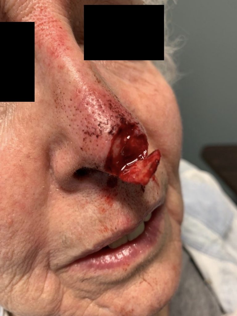
Figure 1. Nasal tip injury upon arrival
Treatment:
After adequate hemostasis and local anesthesia administration, the wound was thoroughly irrigated with saline (approximately 250cc) and removing any foreign debris. The avulsed nasal tip was seemingly adequately perfused and approximated well. Given patient’s preference, 5-0 fast absorbing gut was used in an interrupted fashion approximately 2mm apart. Bacitracin was applied to the wound and the patient was instructed to apply Bacitracin ointment twice daily until follow up. She was also instructed to avoid sun exposure and use SPF-30 or higher lotion if exposed to sun for the next year. This was done to decrease the chance of hyper pigmentation of her scar. She was scheduled for follow up in 1 week. Given mechanism and exposed cartilage, she was given 10 days of clindamycin. In regard to the nasal bone fracture, standard nasal precautions were provided including avoiding nasal trauma.. Over the counter nasal saline solution as needed for any mucus plugging.
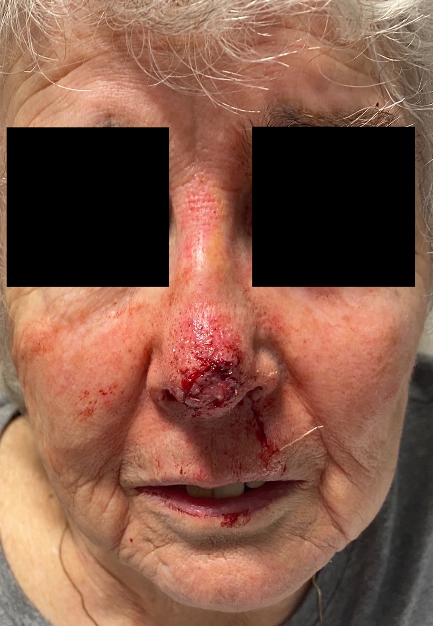
Figure 2. Immediately after repair
Follow Up:
The patient followed up in 2 weeks and was found to have good healing with no signs of infection and minimal scar. Patient instructed to continue using wound care (sunscreen) for at least one year. Patient content with outcome.
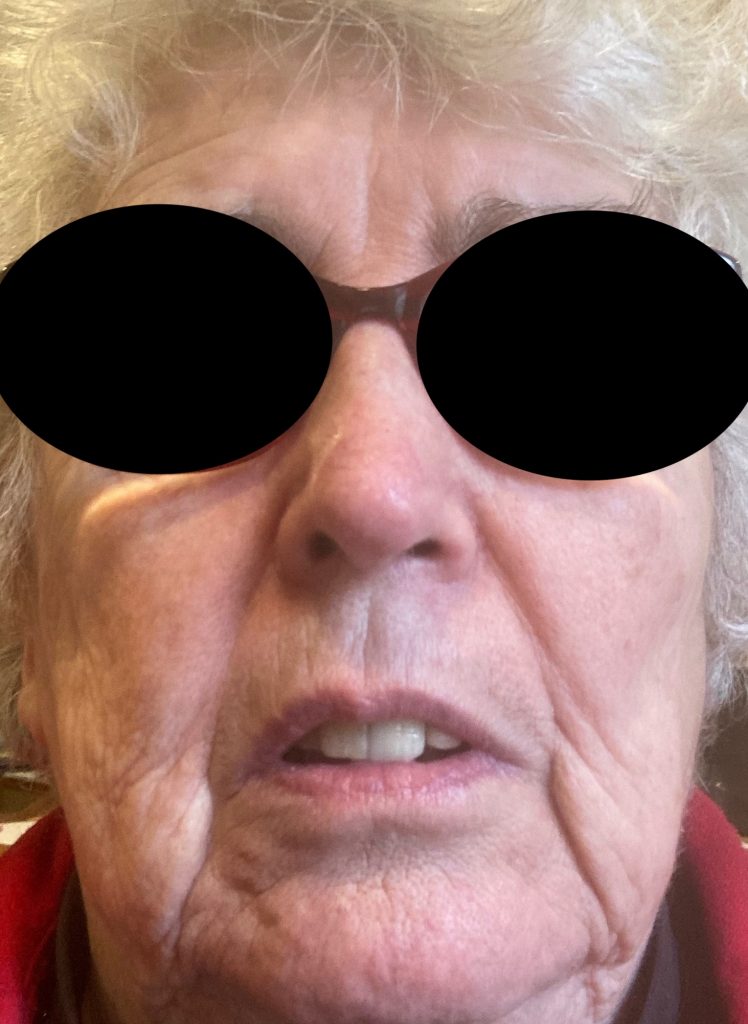
Figure 3. 5 month post repair
