Traumatic Amputation of the Left Index, Middle, and Ring Fingers Following a Lawn Mower Accident

Fortunay Diatta
, Olivier Noel
, Martin Kauke-Navarro
, Omar Allam

History:


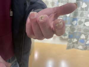
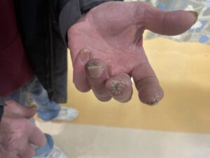
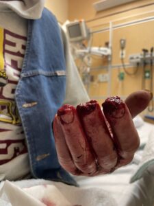
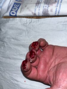
This case report presents a 70-year-old male who sustained amputations of the left index, middle, and ring fingers following a lawn mower accident. He is otherwise healthy.

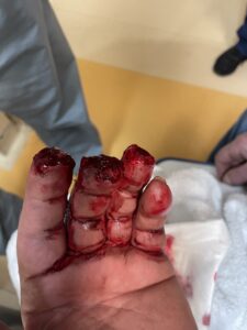
The 3 pictures show the levels of amputation.

Findings:

The x-rays show the level of bony loss in each finger.

of the distal

Upon examination and review of plain X-rays, the following findings are noted:

1. Left index and ring fingers:
– Soft tissue loss at the fingertips with minimal tip fractures and bone loss.
– Minor soft tissue deficits visible, with small osseous fragments at the distal terminus of the residual structures of both digits.

2. Left middle finger:
– Transverse amputation through the base of the distal phalanx, not involving the distal interphalangeal joint.

Upon examination, the left index and ring fingers exhibit minor soft tissue deficits, with visible small osseous fragments at the distal terminus of the residual structures of both digits. The middle finger has undergone a transverse amputation at the base of the distal phalanx. The affected areas are warm with intact blood supply. The distal fingers show a capillary refill time of less than three seconds. The insertion of the flexor digitorum profundus is intact in all 3 injured fingers. Sensory function is intact in the areas innervated by the radial, median, and ulnar nerves.


Diagnosis:

Amputation of tips of left index, middle and ring fingers from lawn more accident. Loss of bone and soft tissue just above the base of the distal phalanx of middle finger and tip soft tissue loss in index and ring with minimal bone loss. Profundus tendon insertion intact in all 3 fingers. No bone, vascular or nerve injury proximal to the amputation.

Differential Diagnoses:

Injuries to the rest of the upper extremities have been ruled out through examination.

Workup Required:

1. X-ray of the left hand to assess the extent of bone injury and guide surgical planning.
2. Complete Blood Count (CBC).

Plan:

1. Administer tetanus prophylaxis to prevent tetanus infection.
2. Initiate intravenous (IV) antibiotics (Ancef) while the patient is in the Emergency Department to prevent infection.
3. Perform surgical revisions of the amputations to shorten exposed bone and cover with soft tissue, minimizing finger shortening as per the patient’s preference.
4. Apply a protective semi-occlusive hand dressing, to be changed every 3-4 days to promote healing and prevent infection.
5. Discharge the patient home with a prescription for oral antibiotics (Keflex) and pain management (Tylenol with codeine) for 5 days.
6. Schedule a follow-up visit in 2 weeks to assess healing and address any complications.


Expertise Needed:

The case requires the expertise of a hand surgeon or plastic surgeon to perform the surgical revisions and manage the patient’s care.

Treatment:

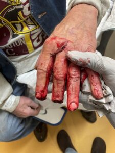
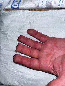
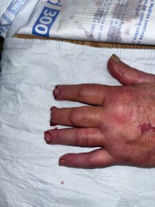
After administering digital blocks to anesthetize the index, middle, and ring fingers, the surgical site was thoroughly irrigated with sterile saline solution. Conservative debridement was performed to remove any devitalized or fragmented tissue, ensuring optimal conditions for wound healing. Primary closure of the skin was achieved over the index and ring fingers; however, a small soft tissue defect remained over the exposed bone of the middle finger. The decision was made to allow this area to heal by secondary intention.

A semi-occlusive dressing (Tegaderm) was applied to protect the surgical site and promote a moist wound healing environment [3]. The patient was discharged with a 5-day course of oral antibiotics (Cephalexin) to prevent infection and Tylenol with codeine for pain management. Close follow-up was arranged to monitor the progress of wound healing and address any potential complications.

Pictures immediately postop.

Follow Up:

At the 2-week follow-up, the patient was found to be healing without any signs of complications. During the 2-month follow-up, the index and ring fingers were noted to be well-healed. The middle finger had a scab over the tip. The possibility of a revision surgery for the middle finger was discussed, but the patient declined the option.
Print Friendly, PDF & Email

References

Sadoma BR, Sheets NW, Plurad DS, Dubina ED. Traumatic Amputations Treated in US Emergency Departments: A Review of the NEISS Database. Am Surg. 2023 Oct;89(10):4123-4128. doi: 10.1177/00031348231177947. Epub 2023 May 24. PMID: 37226454.
Peterson SL, Peterson EL, Wheatley MJ. Management of fingertip amputations. J Hand Surg Am. 2014 Oct;39(10):2093-101. doi: 10.1016/j.jhsa.2014.04.025. PMID: 25257490.
Boudard, J., Loisel, F., El Rifaï, S., Feuvrier, D., Obert, L., & Pluvy, I. (2019). Fingertip amputations treated with occlusive dressings. Hand Surgery and Rehabilitation, 38(4), 257-261
https://doi-org.yale.idm.oclc.org/10.1177/00031348231177947
https://www-sciencedirect-com.yale.idm.oclc.org/science/article/pii/S0363502314005012?via%3Dihub
https://www.sciencedirect.com/science/article/abs/pii/S2468122919300787

Related Articles