Findings:
33 year old woman who had lost her entire scalp and forehead skin to a horizontal line below her eyebrows. Her ears were intact. There was very slight diffuse bleeding from her scalp wound. Her vision and hearing appeared normal.
Physical examination was otherwise normal and her blood pressure is 125/80 and her pulse is 76. An acutely processed blood sample revealed a hematocrit of 37. There was no evidence of any fractures.
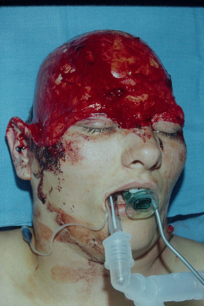
Fig. 1. Shows anterior level of scalp avulsion below eyebrows and across glabella.
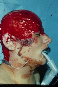
Fig. 2. Shows lateral level of scalp separation.
Treatment:
The patient was acutely taken to the OR and prepared for the procedure. In the meantime the scalp was shaved. Three pairs of arteries and veins were identified; bilateral temporal ones and one supraorbital. A total of 6 vein grafts were used. The frontal branches of the 7th nerve were not found. At the end of the procedure, the scalp was well perfused.
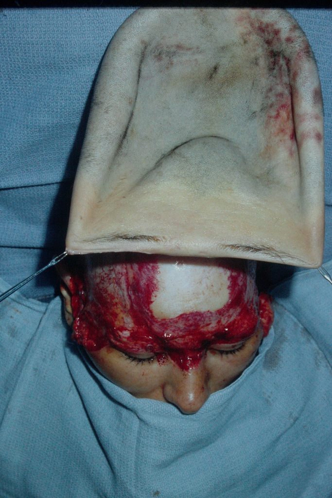
Fig. 3. Scalp ready to be re-attached.
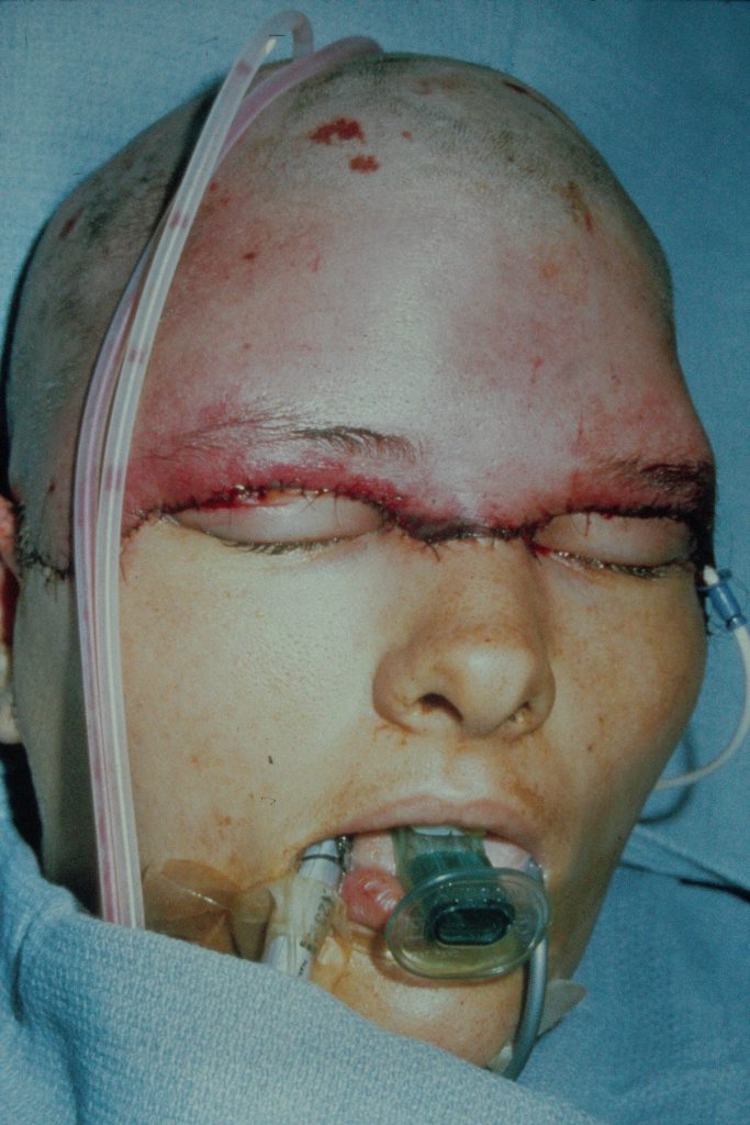
fig. 4. Scalp revascularized with microsurgery. 3 arteries and 3 veins have been reconnected with vein grafts.
Follow Up:
More than 95% of the scalp survived with a minor loss in the neck. The hair growth returned within days. Due to eyebrow ptosis from the denervation of the frontalis muscle, the eyebrows were elevated by bilateral limited full thickness skin excisions. The patient was very happy with the outcome of the procedures.
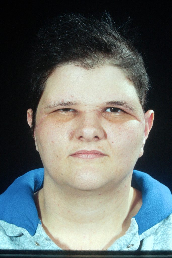
Fig. 5. Healing after 6 months. there is good hair growth but the eyebrows are ptotic.
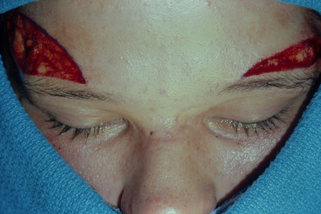
Fig. 6. Bilateral lenticular excisions have been done to raise the eyebrows.
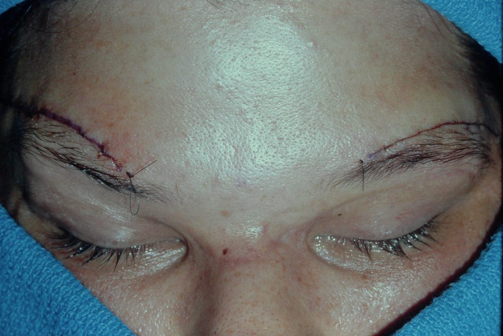
Fig. 7. The excisions have been closed.
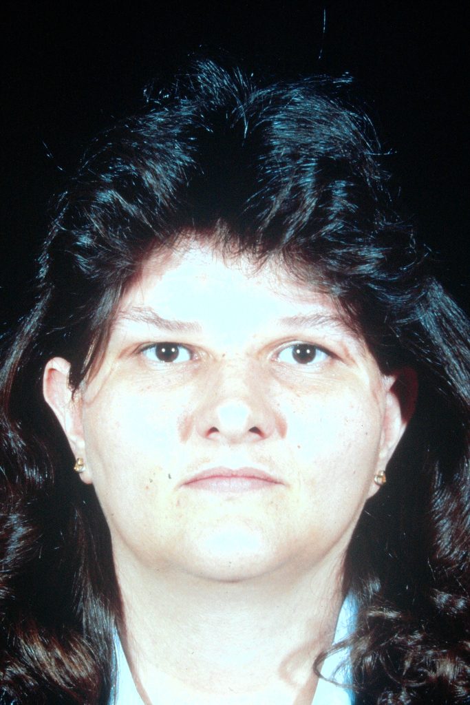
Fig. 8. Final result after 2 years. The patient was extremely pleased with the result.
