History:
The patient is a 28-year-old female who presented to our clinic to improve the appearance of the scar of the philtrum and the upper lip. She experienced a motorcycle accident 8 years before the presentation outside the US and landed with her face on concrete, sustaining multiple facial lacerations and abrasions, including on the upper lip. The lacerations were sutured in the emergency department; however, subsequently, she developed hypertrophic scarring that deformed and shortened her philtrum and distorted her upper lip. The scar was not itchy and not painful. She was constantly wearing a face mask, avoiding exposure of her face in public.
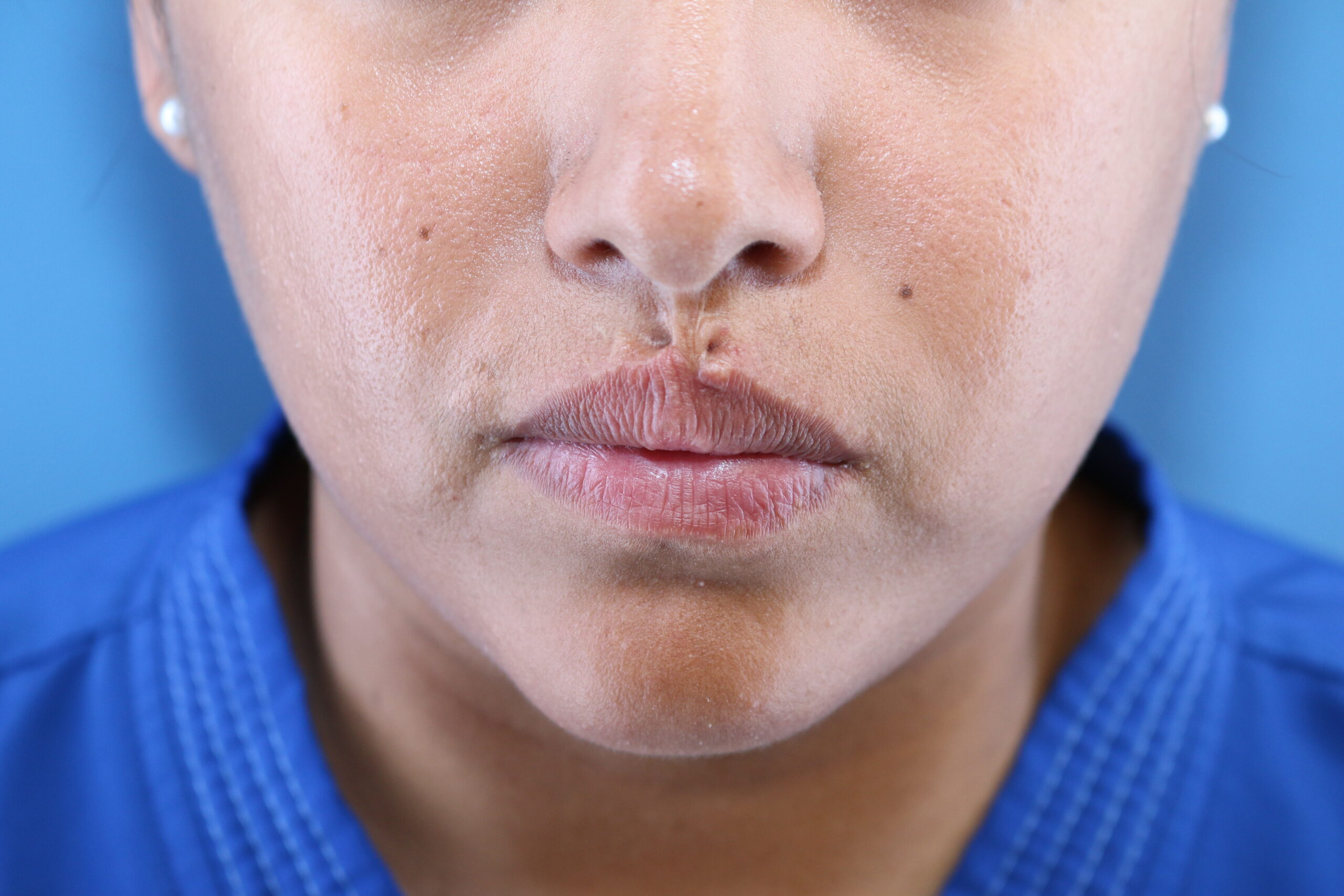
Pre-Operative Assessment of Philtrum scar, anterior view at rest.
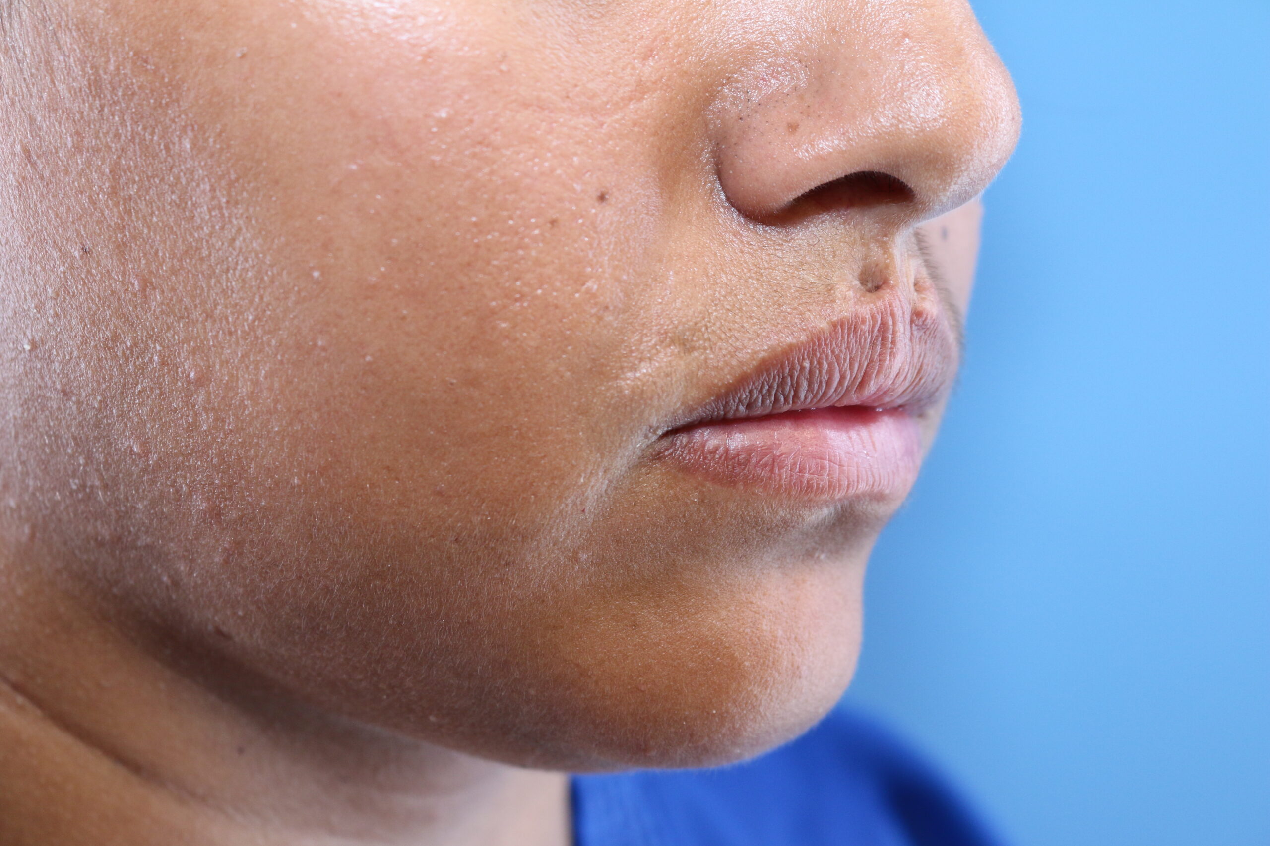
Pre-Operative Assessment of Philtrum scar, right oblique view
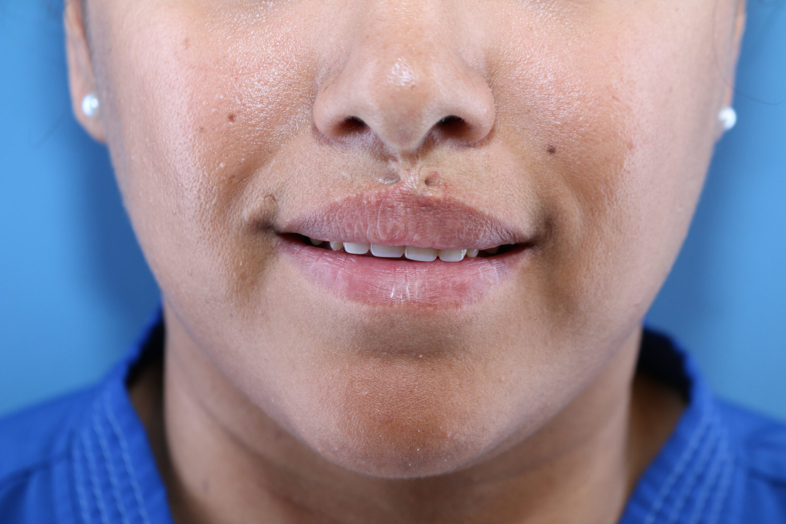
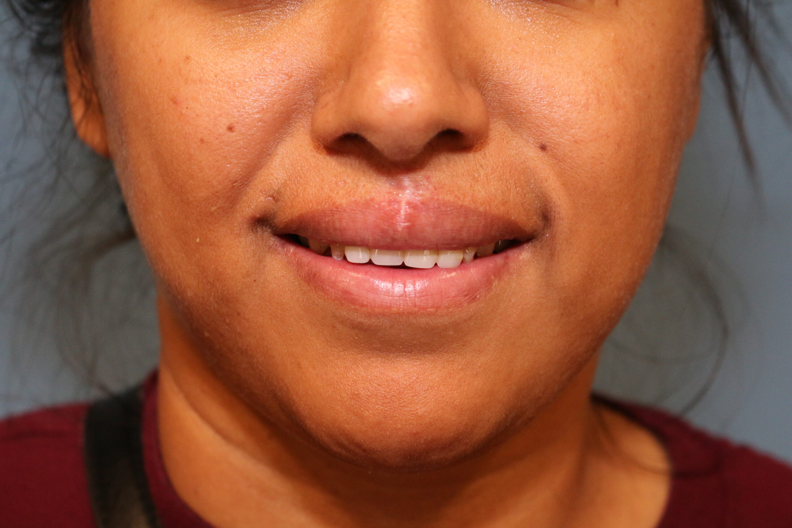
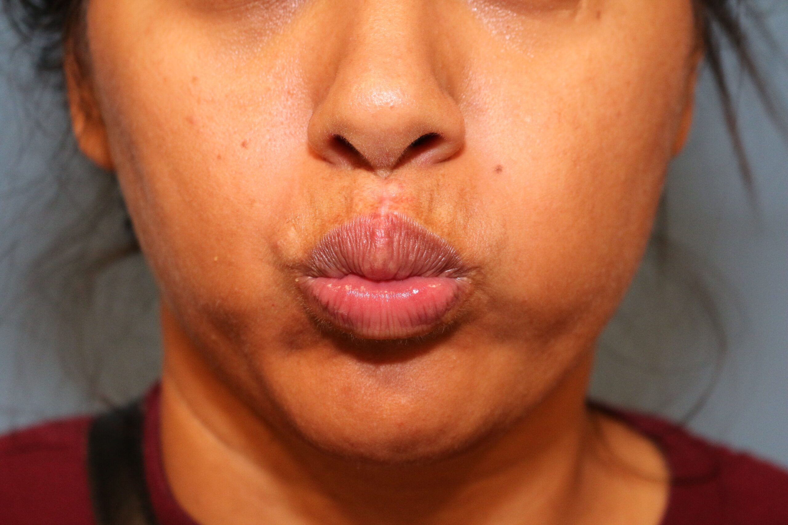
Pre-Operative Assessment of Philtrum scar, anterior view, smiling
Treatment:
The patient was given 10mg of Valium PO, followed by 500mg of Keflex, 30 minutes before incision. An infraorbital block was performed bilaterally with the buccal approach using a total of 3 cc of Lidocaine 1% with epinephrine. The philtrum was infiltrated with Lidocaine 1% with epinephrine; the total required was five cc. The area was then prepared with Betadine x3 and draped in a standard, sterile fashion. The scar bridge was then transected with a #15 blade, and the deformity was evaluated after release. Then, the scar was excised in a rhomboid shape to elongate the philtrum, with the tissue removed all the way down to the orbicularis oris muscle. Undermining of the orbicularis oris was performed medially and laterally 5-10 mm. Two deep 3-0 Vicryl stitch sutures were placed to approximate and plicate the muscle to offload the skin and create a pleasant pi philtral dimple. Bleeding was controlled by electrocautery, pressure, and sutures. Then, deep dermal 5-0 Monocryl sutures were placed, and “dog ears” were excised towards the lip, including vermillion and superiorly towards the columella. And 4-0 chromic gut interrupted sutures were placed superficially on the mucosa, and Prolene 6-0 interrupted skin sutures were placed, starting with re-establishing the vermilion cutaneous border, once completed, Dermabond was applied as a final layer to complete the 4-layered closure. Steristrips were also applied to take the tension off even more. The procedure was tolerated well with no immediate complications, although moderate bleeding was noted. Patient was prescribed 3 days of Keflex and recommended a soft diet to avoid excessive chewing or excessively moving her lips.
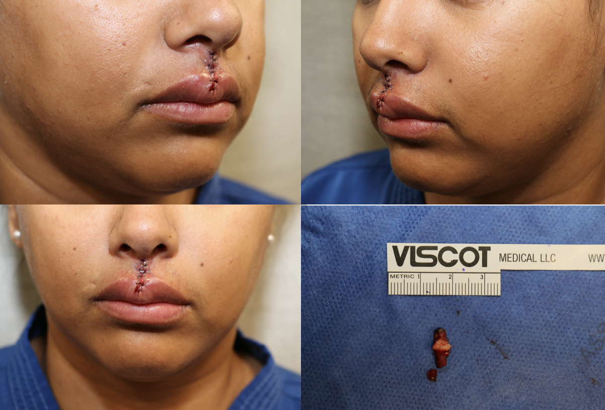
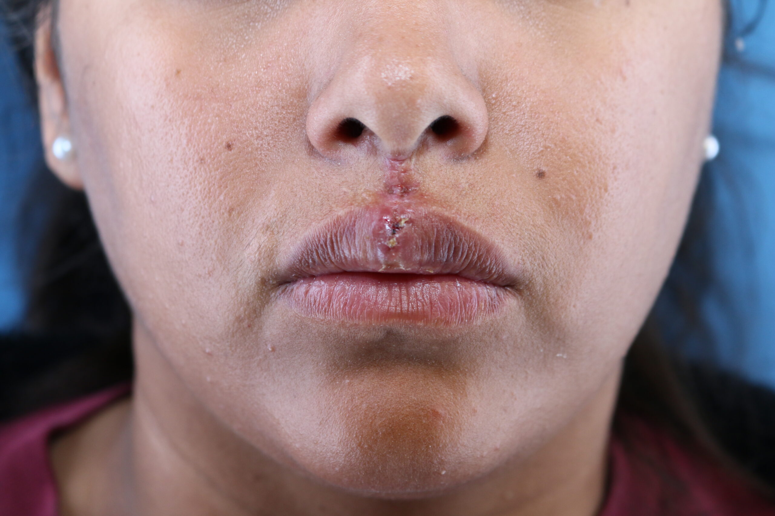
Immediately post-operative pictures after the scar tissue had been excised and the lip reconstructed.
Follow Up:
The first follow-up was at one week after the procedure; permanent sutures were removed, and Dermabond was applied. Good alignment of the cutaneous vermillion border was noticed; the incision was healing well with moderate swelling of the lip.
2nd follow-up was at 3 weeks after the procedure. She was overall doing well; the scar had started becoming more prominent, and 0.5 cc of Kenalog-40 was injected.
At 6 weeks after the procedure, scar tissue was forming, and one deep Vicryl suture was extruding. We removed the suture and instructed the patient to continue deep scar massage, once closed, to continue applying Biocorneum with sunscreen, and plan to follow up to inject more steroid potentially.
At 10 weeks after the procedure, the scar was maturing. We repeated the steroid injection of 0.2 cc Kenalog-40 with the plan to continue massaging the scar and taping (using a silicone sheath nightly). The patient was happy with the results and did not have to wear a covering mask on the face. She was told that a revision procedure might improve the scar but she did not want to pursue this option.
Post-Operative review at 1 week.
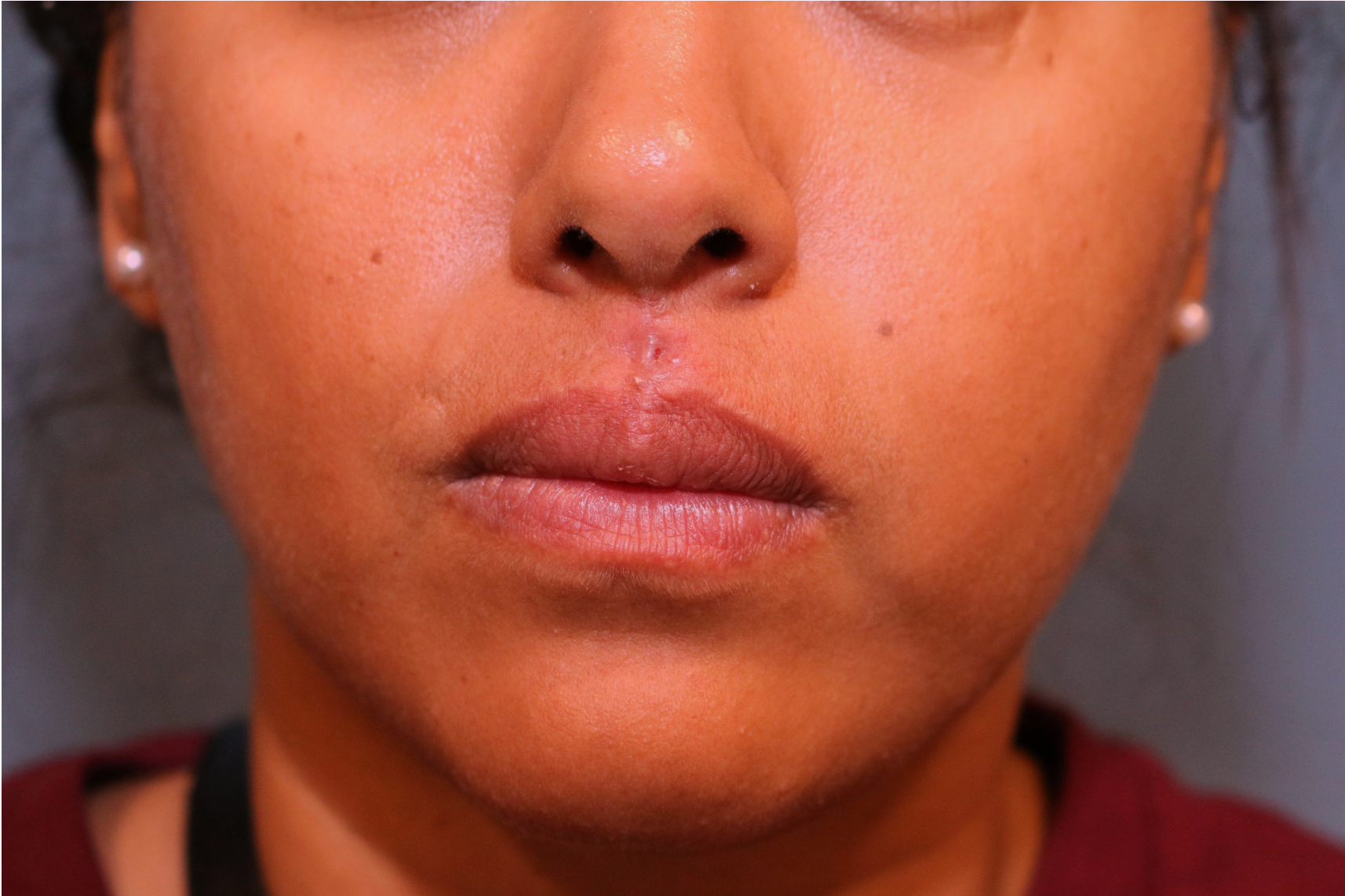
Post-Operative review at 6 weeks.
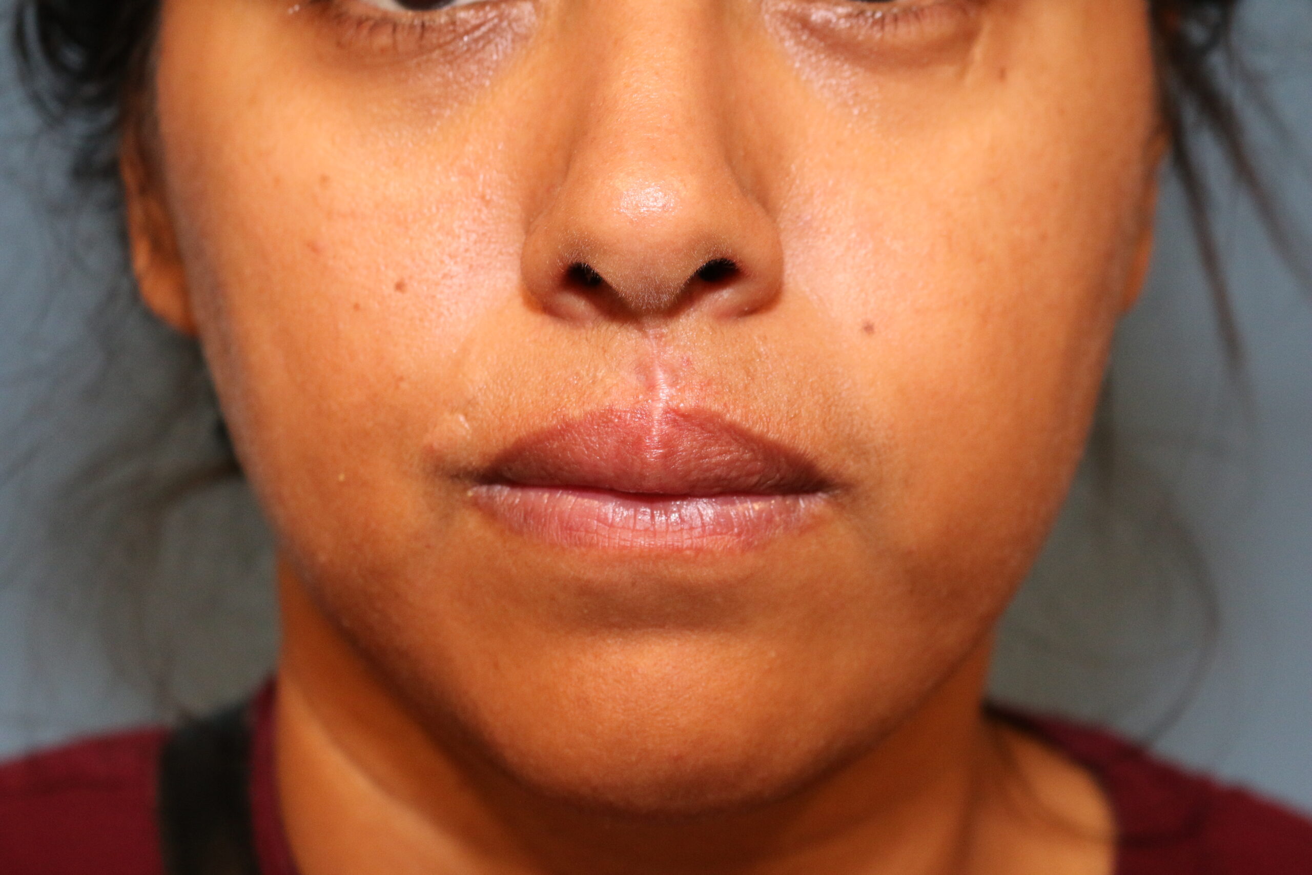
Post-Operative review at 10 weeks anterior view at rest.
Post-Operative review at 10 weeks anterior view at rest.
Post-Operative review at 10 weeks anterior view at rest.
