Infected Index Finger Wound

Jhade Woodall, MD

History:

The patient is a 63 year old farmer who is referred from the ER with a left index finger infection. Five days earlier he tore up a dorsal wound on this finger on a barb wire. Two days ago, he was seen in the ER and started on antibiotics. The finger has become more swollen and painful and when seen in the ER today he is referred to hand surgery. Outside of the current problem, the patient is healthy and he takes no medications.

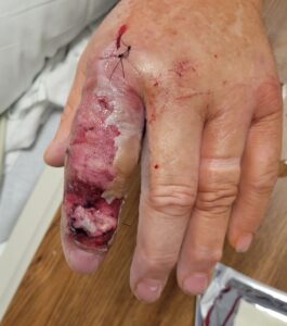
Fig.1. Infected wound on dorsum of index finger.

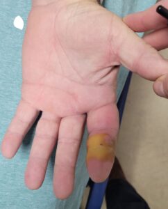
Fig.2. Volar side of infected index finger.

Findings:

Over the DIP joint of the left index finger there is a 14 X 15 mm full thickness wound and partial thickness skin loss over the dorsum of the rest of the finger.. The DIP joint is stiff and has no extension of the distal phalanx. On the volar side of the DIP joint there is an area 22 X 17 mm which is yellow, swollen and with no capillary refill. There is fusiform swelling and tenderness of the whole finger. There is no axillary lymphadenopathy and the patient no fever.

Diagnosis:

Infected left index finger with dorsal and possibly volar skin loss. There is involvement of the DIP joint and the extensor tendon has ruptured. R/O flexor tendon tenosynovitis. No evidence of infection beyond the finger.

Differential Diagnoses:

Osteomyelitis, tenosynovitis.


Workup Required:

Xray of left hand, wound cultures (likely to be negative because of ongoing antibiotic therapy). CBC and electrolytes as per anesthesia.

Plan:

The patient was prepared for surgery under axillary anesthesia with debridement of necrotic tissue, drainage of the subcutaneous space and exploration of the flexor tendons.

Expertise Needed:

Hand Surgeon, plastic or orthopedic.

Fig.3. Dorsal infected wound at time of drainage and debridement.

Fig.4. Exploration of flexor tendons.

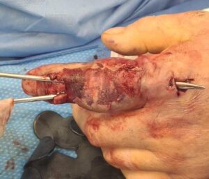
Fig 5. Drainage of dorsal subcutaneous infection.

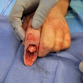
Fig.6. At amputation of distal phalanx, showing volar skin flap and bone of distal middle phalanx.

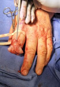
Fig.7. Volar flap covering bone of middle phalanx.

Treatment:

In the OR the wounds were debrided. The flexor tendons were explored and tendon sheaths were opened and showed no evidence of tenosynovitis. Drains were placed in the wounds. The patient was continued on antibiotics. Two weeks later, the infection had cleared. He had no active extension of the DIP joint. The possibilities of DIP joint fusion and flap coverage were discussed as well as an amputation at the DIP level. The patient was greatly in favor of an amputation, which was done shortly thereafter.


Follow Up:

Two months later the wound was completely healed without any remaining infection and the patient had full function of the remaining digit.
Print Friendly, PDF & Email

References

Hand Infections Wendy Z.W. Teo, BA (Cantab), BM BCh (Oxon), LLM Kevin C. Chung, MD, MS
Repair and Reconstruction of Thumb and Finger Tip Injuries: A Global View Clinics in Plastic SurgeryVol. 41Issue 3p325–359Published in issue: July, 2014 Jin Bo TangDavid ElliotRoberto AdaniMichel Saint-CyrFelix Stang
Finger reconstruction with dorsal metacarpal artery perforator flaps and dorsal finger perforator flaps based on the dorsal branches of the palmar digital arteries - 40 consecutive cases Inga S Besmens 1, Marco Guidi 1, Florian S Frueh 1, Semra Uyulmaz 1, Nicole Lindenblatt 1, Lisa Reissner 2, Maurizio Calcagni 1
https://doi.org/10.1016/j.cps.2019.03.003
https://doi.org/10.1016/j.cps.2014.04.004
https://doi.org/10.1080/2000656X.2020.1762624

Related Articles