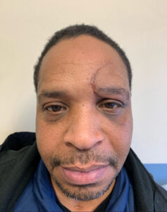
 A 35-year-old male presented to the emergency department with a forehead laceration extending to the upper eyelid. The patient reported sustaining the injury after a fall during a recreational sports activity. No loss of consciousness was reported, and the patient denied any visual disturbances or other associated symptoms.
Treatment:
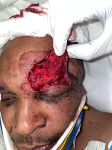
The laceration repair was performed following standard protocol. After obtaining informed consent, the wound was thoroughly explored, and no foreign bodies were identified. The area was then prepped and draped in a sterile fashion. Local anesthesia was administered using 15cc of 1% lidocaine with 1:100,000 epinephrine to ensure patient comfort during the procedure.
Once adequate anesthesia was achieved, a thorough washout of the wound was performed using 2 liters of dilute betadine to minimize the risk of infection. The muscle layer was then carefully approximated and closed using 4-0 Vicryl sutures to ensure proper alignment and support of the deeper tissues.
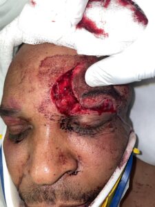
Following the muscle layer closure, attention was turned to the deep dermal layer. This layer was meticulously approximated using 4-0 Monocryl sutures, which provided additional support and improved the cosmetic outcome by reducing tension on the skin surface.
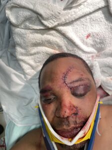
Finally, the skin was closed using 5-0 Prolene interrupted sutures. This technique allowed for precise edge alignment and optimal wound eversion, which is crucial for minimizing scar formation, especially in a cosmetically sensitive area like the forehead and upper eyelid [2,3].
Upon completion of the suturing, a sterile dressing was applied to protect the wound. The patient tolerated the entire procedure well, and no complications were encountered during the repair process.
Follow Up: