Forearm Avulsion Wound
History:
A 68-year-old male with a PMH significant for insulin-dependent type II diabetes presented to the emergency department with a large cutaneous defect of the left upper extremity. The night before, the patient reported falling over his dog into an antique glass cabinet, sustaining multiple lacerations with significant avulsion of the soft tissue of his left forearm. The patient stated that he had removed part of the avulsed tissue flap himself with scissors.

Fig.1. Forearm wound at presentation
Findings:
The patient was in no acute distress on presentation and vital signs were within normal limits. There was an approximately 12 x 15 cm avulsion wound of the soft tissue of the proximal aspect of the left lateral forearm. A small piece of avulsed skin flap remained at the proximal aspect of the wound, but the portion of the avulsed soft tissue distally had been sharply removed by the patient. The wound was not actively bleeding. Distal pulses of the left upper extremity were intact with brisk capillary refill. Motor and sensory function were intact distal to the wound.
Diagnosis:
Differential Diagnoses:
There was some concern initially for neurovascular injury, but physical exam was reassuring with intact pulses and normal neurological function distal to the injury.
Workup Required:
BMP and CBC were ordered. Most recent albumin and HbA1c were 4.5 and 9.6% respectively.
Plan:
The patient had already been given a dose of TDap and Cefazolin prior to plastic surgery consultation. The plan was for the patient to be taken to the operating room for irrigation and debridement of the wound under general anesthesia, with closure of the wound with a split-thickness skin graft, pressure dressing. Follow-up would be scheduled for 5 days post-op in clinic.
Expertise Needed:
Plastic surgeon or Hand surgeon or Surgeon with skin grafting experience
Treatment:
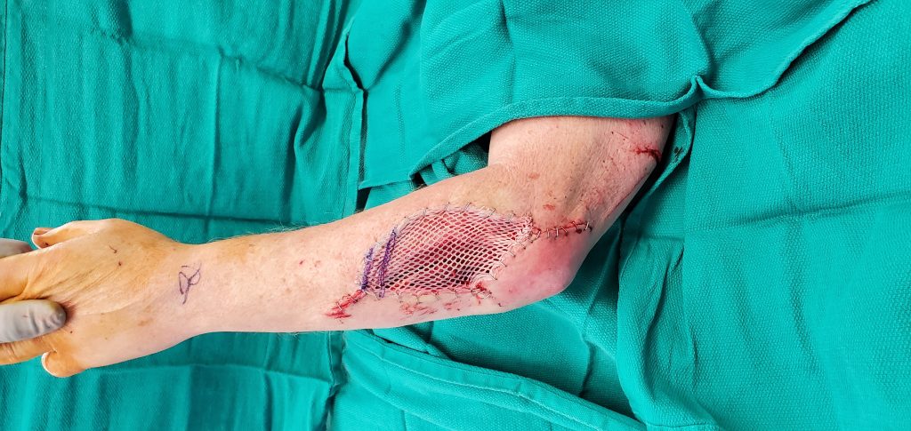
The patient was taken to the operating room for irrigation and debridement of his forearm wound under general anesthesia. Proximally, primary closure was achieved with buried Monocryl sutures along an approximately 5 cm segment the wound. The remaining defect, approximately 7 x 12 cm, was covered with a split-thickness skin graft taken from the left anterior thigh with 3-inch dermatome blade, 0.016 inch graft thickness. The donor site was initially marked and pre-injected with a 50:50% mix of Lidocaine 1% with 1:100000 epinephrine and Marcaine 0.25% to a total of 20 ml. The graft was meshed 1.5:1 and secured to the forearm with staples. The recipient site was dressed with Xeroform, wet to dry Kerlix gauze and secured with an Ace wrap. The donor site was dressed with one layer of Xeroform, 3M foam and ACE wrap.

Fig.2. Split-thickness skin graft to the forearm avulsion wound
Follow Up:
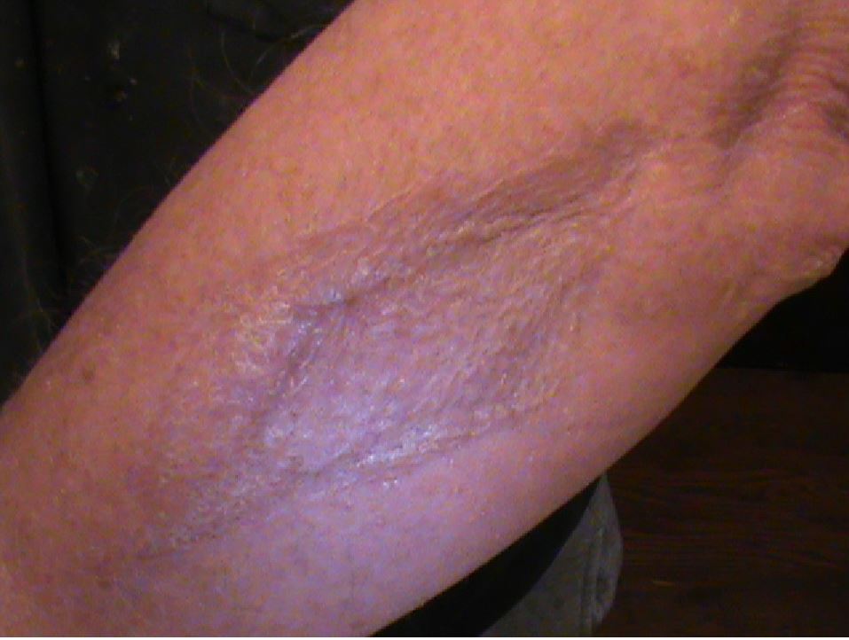
Patient was seen in clinic 5 days post-operatively, on exam the graft appeared viable with good take with no signs of infection, minor swelling in his left hand was noted. At 2 weeks post-op, the swelling had resolved, and the graft continued to look healthy and viable with excellent take, at that time staples were removed.

Fig.3. Forearm wound at 1-year follow up

