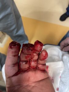
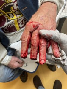
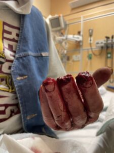
Traumatic Amputation of the Left Index, Middle, and Ring Fingers Following a Lawn Mower Accident
Findings:

The x-rays show the level of bony loss in each finger.
of the distal

Upon examination and review of plain X-rays, the following findings are noted:
1. Left index and ring fingers:
– Soft tissue loss at the fingertips with minimal tip fractures and bone loss.
– Minor soft tissue deficits visible, with small osseous fragments at the distal terminus of the residual structures of both digits.
2. Left middle finger:
– Transverse amputation through the base of the distal phalanx, not involving the distal interphalangeal joint.
Upon examination, the left index and ring fingers exhibit minor soft tissue deficits, with visible small osseous fragments at the distal terminus of the residual structures of both digits. The middle finger has undergone a transverse amputation at the base of the distal phalanx. The affected areas are warm with intact blood supply. The distal fingers show a capillary refill time of less than three seconds. The insertion of the flexor digitorum profundus is intact in all 3 injured fingers. Sensory function is intact in the areas innervated by the radial, median, and ulnar nerves.
Diagnosis:
Differential Diagnoses:
Workup Required:
2. Complete Blood Count (CBC).
Plan:
1. Administer tetanus prophylaxis to prevent tetanus infection.
2. Initiate intravenous (IV) antibiotics (Ancef) while the patient is in the Emergency Department to prevent infection.
3. Perform surgical revisions of the amputations to shorten exposed bone and cover with soft tissue, minimizing finger shortening as per the patient’s preference.
4. Apply a protective semi-occlusive hand dressing, to be changed every 3-4 days to promote healing and prevent infection.
5. Discharge the patient home with a prescription for oral antibiotics (Keflex) and pain management (Tylenol with codeine) for 5 days.
6. Schedule a follow-up visit in 2 weeks to assess healing and address any complications.
Expertise Needed:
Treatment:
After administering digital blocks to anesthetize the index, middle, and ring fingers, the surgical site was thoroughly irrigated with sterile saline solution. Conservative debridement was performed to remove any devitalized or fragmented tissue, ensuring optimal conditions for wound healing. Primary closure of the skin was achieved over the index and ring fingers; however, a small soft tissue defect remained over the exposed bone of the middle finger. The decision was made to allow this area to heal by secondary intention.
A semi-occlusive dressing (Tegaderm) was applied to protect the surgical site and promote a moist wound healing environment [3]. The patient was discharged with a 5-day course of oral antibiotics (Cephalexin) to prevent infection and Tylenol with codeine for pain management. Close follow-up was arranged to monitor the progress of wound healing and address any potential complications.
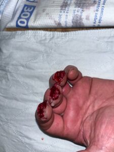
Pictures immediately postop.
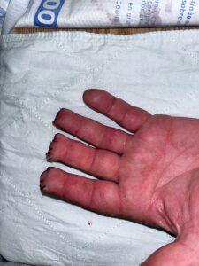
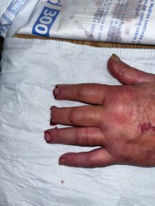
Follow Up:

